gamma-delta T cell activation / NK T cell differentiation / activation of phospholipase C activity / Generation of second messenger molecules / cellular defense response / T cell activation / FCERI mediated Ca+2 mobilization / positive regulation of cytokine production / B cell receptor signaling pathway / non-specific protein-tyrosine kinase ...gamma-delta T cell activation / NK T cell differentiation / activation of phospholipase C activity / Generation of second messenger molecules / cellular defense response / T cell activation / FCERI mediated Ca+2 mobilization / positive regulation of cytokine production / B cell receptor signaling pathway / non-specific protein-tyrosine kinase / non-membrane spanning protein tyrosine kinase activity / cell-cell junction / T cell receptor signaling pathway / adaptive immune response / intracellular signal transduction / phosphorylation / signal transduction / ATP binding / metal ion binding / nucleus / plasma membrane / cytosol Similarity search - Function
Resolution: 1.71→50.6 Å / Cor.coef. Fo:Fc: 0.965 / Cor.coef. Fo:Fc free: 0.944 / SU B: 4.856 / SU ML: 0.079 Isotropic thermal model: individual atomic plus TLS in one group Cross valid method: THROUGHOUT / σ(F): 0 / ESU R: 0.108 / ESU R Free: 0.108 / Stereochemistry target values: MAXIMUM LIKELIHOOD / Details: HYDROGENS HAVE BEEN ADDED IN THE RIDING POSITIONS
Rfactor
Num. reflection
% reflection
Selection details
Rfree
0.20164
1295
5.1 %
RANDOM
Rwork
0.15906
-
-
-
obs
0.16125
24162
99.15 %
-
all
-
25163
-
-
Solvent computation
Ion probe radii: 0.8 Å / Shrinkage radii: 0.8 Å / VDW probe radii: 1.2 Å / Solvent model: MASK
 Movie
Movie Controller


 Open data
 Basic information
 Components
 Keywords
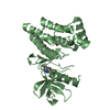
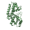
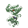
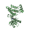
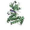
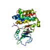
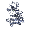
 protein kinase / phospho-transfer / TRANSFERASE-TRANSFERASE INHIBITOR complex
 Function and homology information
 Authors
 Citation
 Structure visualization
 Downloads & links
 Other downloads
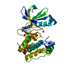


 PDBj
PDBj


 Assembly
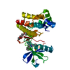


 Mass: 420.550 Da / Num. of mol.: 1 / Source method: obtained synthetically / Formula: C24H32N6O
 Mass: 162.227 Da / Num. of mol.: 1 / Source method: obtained synthetically / Formula: C8H18O3
 Mass: 18.015 Da / Num. of mol.: 222 / Source method: isolated from a natural source / Formula: H2O
 Sample preparation
 / Beamline: I04 / Wavelength: 0.9795 Å
 Processing