gamma-delta T cell activation / NK T cell differentiation / activation of phospholipase C activity / Generation of second messenger molecules / cellular defense response / T cell activation / FCERI mediated Ca+2 mobilization / positive regulation of cytokine production / B cell receptor signaling pathway / non-specific protein-tyrosine kinase ...gamma-delta T cell activation / NK T cell differentiation / activation of phospholipase C activity / Generation of second messenger molecules / cellular defense response / T cell activation / FCERI mediated Ca+2 mobilization / positive regulation of cytokine production / B cell receptor signaling pathway / non-specific protein-tyrosine kinase / non-membrane spanning protein tyrosine kinase activity / cell-cell junction / T cell receptor signaling pathway / adaptive immune response / intracellular signal transduction / phosphorylation / signal transduction / ATP binding / metal ion binding / nucleus / plasma membrane / cytosol Similarity search - Function
 Movie
Movie Controller

 Yorodumi
 Open data
 Basic information
 Components
 Keywords
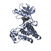
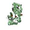
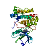
 KINASE DOMAIN / Transferase-Transferase inhibitor complex
 Function and homology information
 Authors
 Citation
 Structure visualization
 Downloads & links
 Other downloads
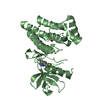


 PDBj
PDBj


 Assembly


 Mass: 466.559 Da / Num. of mol.: 3 / Source method: obtained synthetically / Formula: C22H26N8O2S
 Mass: 468.575 Da / Num. of mol.: 2 / Source method: obtained synthetically / Formula: C22H28N8O2S
 Mass: 96.063 Da / Num. of mol.: 4 / Source method: obtained synthetically / Formula: SO4
 Mass: 92.094 Da / Num. of mol.: 1 / Source method: obtained synthetically / Formula: C3H8O3
 Sample preparation
 / Beamline: I03 / Wavelength: 0.9763 Å
 Processing