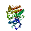
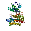
Entry Database : PDB / ID : 4c3fTitle Structure of Lck in complex with a compound discovered by Virtual Fragment Linking TYROSINE-PROTEIN KINASE LCK Keywords Function / homology Function Domain/homology Component
/ / / / / / / / / / / / / / / / / / / / / / / / / / / / / / / / / / / / / / / / / / / / / / / / / / / / / / / / / / / / / / / / / / / / / / / / / / / / / / / / / / / / / / / / / / / / / / / / / / / / / / / / / / / / / / / / / / / / / / / Biological species HOMO SAPIENS (human)Method / / Resolution : 1.72 Å Authors Cowan-Jacob, S.W. / Rummel, G. / Stark, W. Journal : J. Med. Chem. / Year : 2013Title : Efficient search of chemical space: navigating from fragments to structurally diverse chemotypes.Authors : Wassermann, A.M. / Kutchukian, P.S. / Lounkine, E. / Luethi, T. / Hamon, J. / Bocker, M.T. / Malik, H.A. / Cowan-Jacob, S.W. / Glick, M. History Deposition Aug 23, 2013 Deposition site / Processing site Revision 1.0 Oct 23, 2013 Provider / Type Revision 1.1 Jan 15, 2014 Group Revision 1.2 Dec 14, 2016 Group / OtherRevision 1.3 Apr 4, 2018 Group / Category / Item Revision 1.4 Feb 27, 2019 Group / Database references / Experimental preparationCategory citation / exptl_crystal_grow ... citation / exptl_crystal_grow / pdbx_database_proc / struct_biol Item _citation.journal_abbrev / _citation.journal_id_ISSN ... _citation.journal_abbrev / _citation.journal_id_ISSN / _citation.page_last / _citation.pdbx_database_id_DOI / _citation.title / _exptl_crystal_grow.temp Revision 1.5 Dec 20, 2023 Group Data collection / Database references ... Data collection / Database references / Derived calculations / Other / Refinement description Category chem_comp_atom / chem_comp_bond ... chem_comp_atom / chem_comp_bond / database_2 / pdbx_database_status / pdbx_initial_refinement_model / struct_site Item _database_2.pdbx_DOI / _database_2.pdbx_database_accession ... _database_2.pdbx_DOI / _database_2.pdbx_database_accession / _pdbx_database_status.status_code_sf / _struct_site.pdbx_auth_asym_id / _struct_site.pdbx_auth_comp_id / _struct_site.pdbx_auth_seq_id
Show all Show less
 Movie
Movie Controller

 Yorodumi
 Open data
 Basic information
 Components
 Keywords
 TRANSFERASE
 Function and homology information
 Authors
 Citation
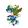
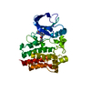
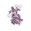
 Structure visualization
 Downloads & links
 Other downloads


 PDBj
PDBj


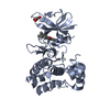
 Assembly


 Mass: 313.356 Da / Num. of mol.: 1 / Source method: obtained synthetically / Formula: C19H15N5
 Mass: 18.015 Da / Num. of mol.: 355 / Source method: isolated from a natural source / Formula: H2O
 Sample preparation
 Processing