 Movie
Movie Controller

[English] 日本語
 Yorodumi
- PDB-4jva: Crystal Structure of RIIbeta(108-402) bound to HE33, a N6 di-prop... -
+ Open data

- Basic information
| Entry | Database: PDB / ID: 4jva | ||||||
|---|---|---|---|---|---|---|---|
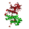
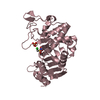
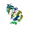
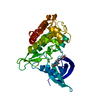
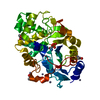
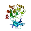
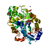
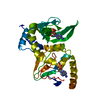
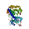
| Title | Crystal Structure of RIIbeta(108-402) bound to HE33, a N6 di-propyl substituted cAMP analog | ||||||
 Components Components | cAMP-dependent protein kinase type II-beta regulatory subunit CAMP-dependent pathway CAMP-dependent pathway | ||||||
 Keywords Keywords | TRANSFERASE/TRANSFERASE INHIBITOR / cAMP-dependent protein kinase / cyclic nucleotide analogs / isoform selectivity / fluorescence anisotropy / TRANSFERASE-TRANSFERASE INHIBITOR complex | ||||||
| Function / homology |  Function and homology information Function and homology informationGPER1 signaling / PKA activation in glucagon signalling / CREB1 phosphorylation through the activation of Adenylate Cyclase / DARPP-32 events / Loss of Nlp from mitotic centrosomes / Recruitment of mitotic centrosome proteins and complexes / Loss of proteins required for interphase microtubule organization from the centrosome / Recruitment of NuMA to mitotic centrosomes / Anchoring of the basal body to the plasma membrane / AURKA Activation by TPX2 ...GPER1 signaling / PKA activation in glucagon signalling / CREB1 phosphorylation through the activation of Adenylate Cyclase / DARPP-32 events / Loss of Nlp from mitotic centrosomes / Recruitment of mitotic centrosome proteins and complexes / Loss of proteins required for interphase microtubule organization from the centrosome / Recruitment of NuMA to mitotic centrosomes / Anchoring of the basal body to the plasma membrane / AURKA Activation by TPX2 / Factors involved in megakaryocyte development and platelet production / Regulation of PLK1 Activity at G2/M Transition / Hedgehog 'off' state / PKA activation / response to antipsychotic drug / cAMP-dependent protein kinase regulator activity / Vasopressin regulates renal water homeostasis via Aquaporins / cAMP-dependent protein kinase inhibitor activity / ciliary base / cAMP-dependent protein kinase complex / protein kinase A catalytic subunit binding / cAMP binding / fatty acid metabolic process / dendritic shaft / learning / regulation of protein phosphorylation / modulation of chemical synaptic transmission / postsynapse / dendritic spine / protein domain specific binding / centrosome / neuronal cell body / dendrite / glutamatergic synapse / ubiquitin protein ligase binding / protein kinase binding / perinuclear region of cytoplasm / plasma membrane / cytosol / cytoplasmSimilarity search - Function | ||||||
| Biological species |  Rattus norvegicus (Norway rat) Rattus norvegicus (Norway rat) | ||||||
| Method | X-RAY DIFFRACTION / SYNCHROTRON / MOLECULAR REPLACEMENT / Resolution: 2.5 Å | ||||||
 Authors Authors | Brown, S.H.J. / Cheng, C.Y. / Saldanha, A.S. / Wu, J. / Cottam, H. / Sankaran, B. / Taylor, S.S. | ||||||
 Citation Citation | Journal: Acs Chem.Biol. / Year: 2013 Title: Implementing Fluorescence Anisotropy Screening and Crystallographic Analysis to Define PKA Isoform-Selective Activation by cAMP Analogs. Authors: Brown, S.H. / Cheng, C.Y. / Saldanha, S.A. / Wu, J. / Cottam, H.B. / Sankaran, B. / Taylor, S.S. | ||||||
| History |
|
- Structure visualization
| Structure viewer | Molecule: MolmilJmol/JSmol |
|---|
- Downloads & links
-Download
| PDBx/mmCIF format | 4jva.cif.gz | 68.7 KB | Display | PDBx/mmCIF format |
|---|---|---|---|---|
| PDB format | pdb4jva.ent.gz | 48.8 KB | Display | PDB format |
| PDBx/mmJSON format | 4jva.json.gz | Tree view | PDBx/mmJSON format | |
| Others |  Other downloads Other downloads |
-Validation report
| Arichive directory | https://data.pdbj.org/pub/pdb/validation_reports/jv/4jvaftp://data.pdbj.org/pub/pdb/validation_reports/jv/4jva | HTTPS FTP |
|---|
-Related structure data
| Related structure data | 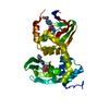 4jv4C  1cx4S S: Starting model for refinement C: citing same article ( |
|---|---|
| Similar structure data |

-Links
 PDBj
PDBj


- Assembly
| Deposited unit | 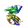
| ||||||||
|---|---|---|---|---|---|---|---|---|---|
| 1 |
| ||||||||
| Unit cell |
|
-Components
| #1: Protein | CAMP-dependent pathway Mass: 34157.719 Da / Num. of mol.: 1 / Fragment: RIIbeta(108-402) of cAMP-dependent Protein Kinase / Mutation: deletion mutant Source method: isolated from a genetically manipulated source Source: (gene. exp.) Rattus norvegicus (Norway rat) / Gene: Prkar2b / Production host:  Escherichia coli (E. coli) / References: UniProt: P12369 Escherichia coli (E. coli) / References: UniProt: P12369 | ||
|---|---|---|---|
| #2: Chemical |   Mass: 413.365 Da / Num. of mol.: 2 / Source method: obtained synthetically / Formula: C16H24N5O6P Mass: 413.365 Da / Num. of mol.: 2 / Source method: obtained synthetically / Formula: C16H24N5O6P#3: Water | ChemComp-HOH / | Water Mass: 18.015 Da / Num. of mol.: 71 / Source method: isolated from a natural source / Formula: H2O Mass: 18.015 Da / Num. of mol.: 71 / Source method: isolated from a natural source / Formula: H2O |
-Experimental details
-Experiment
| Experiment | Method: X-RAY DIFFRACTION / Number of used crystals: 1 |
|---|
- Sample preparation
| Crystal | Density Matthews: 2.17 Å3/Da / Density % sol: 43.44 % |
|---|---|
| Crystal grow | Temperature: 298 K / Method: vapor diffusion under oil (vduo) / pH: 6 Details: 20% PEG 4000, 80 mM Bis-Tris 6.0, and 50 mM MgCl2 using the Oryx crystallization robot (Douglas Instruments) in modified microbatch mode, vapor diffusion under oil (VDUO) , temperature 298.0K |
-Data collection
| Diffraction | Mean temperature: 100 K |
|---|---|
| Diffraction source | Source: SYNCHROTRON / Site: ALS  / Beamline: 8.2.2 / Wavelength: 1 Å / Beamline: 8.2.2 / Wavelength: 1 Å |
| Detector | Type: ADSC QUANTUM 315 / Detector: CCD / Date: Aug 18, 2010 |
| Radiation | Monochromator: GRAPHITE / Protocol: SINGLE WAVELENGTH / Monochromatic (M) / Laue (L): M / Scattering type: x-ray |
| Radiation wavelength | Wavelength: 1 Å / Relative weight: 1 |
| Reflection | Resolution: 2.47→50 Å / Num. obs: 15916 / % possible obs: 95.1 % / Observed criterion σ(F): 2 / Redundancy: 25.1 % / Rmerge(I) obs: 0.08 / Net I/σ(I): 39.3 |
| Reflection shell | Resolution: 2.47→2.58 Å / Redundancy: 19.1 % / Rmerge(I) obs: 0.325 / Mean I/σ(I) obs: 5.5 / % possible all: 88.4 |
- Processing
| Software |
| ||||||||||||||||||||||||||||||||||||||||||||||||||||||||||||
|---|---|---|---|---|---|---|---|---|---|---|---|---|---|---|---|---|---|---|---|---|---|---|---|---|---|---|---|---|---|---|---|---|---|---|---|---|---|---|---|---|---|---|---|---|---|---|---|---|---|---|---|---|---|---|---|---|---|---|---|---|---|
| Refinement | Method to determine structure: MOLECULAR REPLACEMENT Starting model: PDB ENTRY 1CX4 Resolution: 2.5→50 Å / Isotropic thermal model: ANISOTROPIC / Cross valid method: THROUGHOUT / σ(F): 2 / Stereochemistry target values: Engh & Huber
| ||||||||||||||||||||||||||||||||||||||||||||||||||||||||||||
| Displacement parameters |
| ||||||||||||||||||||||||||||||||||||||||||||||||||||||||||||
| Refinement step | Cycle: LAST / Resolution: 2.5→50 Å
| ||||||||||||||||||||||||||||||||||||||||||||||||||||||||||||
| Refine LS restraints |
|
