 Movie
Movie Controller

[English] 日本語
 Yorodumi
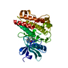
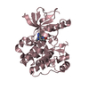
- PDB-1irk: CRYSTAL STRUCTURE OF THE TYROSINE KINASE DOMAIN OF THE HUMAN INSU... -
+ Open data

- Basic information
| Entry | Database: PDB / ID: 1irk | ||||||
|---|---|---|---|---|---|---|---|
| Title | CRYSTAL STRUCTURE OF THE TYROSINE KINASE DOMAIN OF THE HUMAN INSULIN RECEPTOR | ||||||
 Components Components | INSULIN RECEPTOR TYROSINE KINASE DOMAIN | ||||||
 Keywords Keywords | TRANSFERASE (PHOSPHOTRANSFERASE) | ||||||
| Function / homology |  Function and homology information Function and homology informationregulation of female gonad development / positive regulation of meiotic cell cycle / positive regulation of developmental growth /  insulin-like growth factor II binding / male sex determination / exocrine pancreas development / insulin receptor complex / insulin-like growth factor I binding / insulin receptor activity / positive regulation of protein-containing complex disassembly ...regulation of female gonad development / positive regulation of meiotic cell cycle / positive regulation of developmental growth / insulin-like growth factor II binding / male sex determination / exocrine pancreas development / insulin receptor complex / insulin-like growth factor I binding / insulin receptor activity / positive regulation of protein-containing complex disassembly / cargo receptor activity / dendritic spine maintenance / insulin binding / PTB domain binding / adrenal gland development / neuronal cell body membrane / Signaling by Insulin receptor / IRS activation / activation of protein kinase activity / amyloid-beta clearance / positive regulation of respiratory burst / regulation of embryonic development / positive regulation of receptor internalization / transport across blood-brain barrier / insulin receptor substrate binding / positive regulation of glycogen biosynthetic process / epidermis development / Signal attenuation / phosphatidylinositol 3-kinase binding / heart morphogenesis / dendrite membrane / Insulin receptor recycling / insulin-like growth factor receptor binding / neuron projection maintenance / positive regulation of glycolytic process / activation of protein kinase B activity / positive regulation of mitotic nuclear division / receptor-mediated endocytosis / Insulin receptor signalling cascade / learning / caveola / positive regulation of glucose import / positive regulation of MAP kinase activity / receptor internalization / receptor protein-tyrosine kinase / memory / cellular response to growth factor stimulus / peptidyl-tyrosine phosphorylation / cellular response to insulin stimulus / positive regulation of nitric oxide biosynthetic process / male gonad development / late endosome / insulin receptor signaling pathway / glucose homeostasis / amyloid-beta binding / PI5P, PP2A and IER3 Regulate PI3K/AKT Signaling / protein tyrosine kinase activity / positive regulation of MAPK cascade / protein autophosphorylation / lysosome / positive regulation of phosphatidylinositol 3-kinase/protein kinase B signal transduction / receptor complex / endosome membrane / positive regulation of cell migration / symbiont entry into host cell / positive regulation of protein phosphorylation / G protein-coupled receptor signaling pathway / protein domain specific binding / external side of plasma membrane / axon / protein phosphorylation / positive regulation of cell population proliferation / protein-containing complex binding / regulation of DNA-templated transcription / GTP binding / positive regulation of DNA-templated transcription / extracellular exosome / ATP binding / membrane / identical protein binding / plasma membrane insulin-like growth factor II binding / male sex determination / exocrine pancreas development / insulin receptor complex / insulin-like growth factor I binding / insulin receptor activity / positive regulation of protein-containing complex disassembly ...regulation of female gonad development / positive regulation of meiotic cell cycle / positive regulation of developmental growth / insulin-like growth factor II binding / male sex determination / exocrine pancreas development / insulin receptor complex / insulin-like growth factor I binding / insulin receptor activity / positive regulation of protein-containing complex disassembly / cargo receptor activity / dendritic spine maintenance / insulin binding / PTB domain binding / adrenal gland development / neuronal cell body membrane / Signaling by Insulin receptor / IRS activation / activation of protein kinase activity / amyloid-beta clearance / positive regulation of respiratory burst / regulation of embryonic development / positive regulation of receptor internalization / transport across blood-brain barrier / insulin receptor substrate binding / positive regulation of glycogen biosynthetic process / epidermis development / Signal attenuation / phosphatidylinositol 3-kinase binding / heart morphogenesis / dendrite membrane / Insulin receptor recycling / insulin-like growth factor receptor binding / neuron projection maintenance / positive regulation of glycolytic process / activation of protein kinase B activity / positive regulation of mitotic nuclear division / receptor-mediated endocytosis / Insulin receptor signalling cascade / learning / caveola / positive regulation of glucose import / positive regulation of MAP kinase activity / receptor internalization / receptor protein-tyrosine kinase / memory / cellular response to growth factor stimulus / peptidyl-tyrosine phosphorylation / cellular response to insulin stimulus / positive regulation of nitric oxide biosynthetic process / male gonad development / late endosome / insulin receptor signaling pathway / glucose homeostasis / amyloid-beta binding / PI5P, PP2A and IER3 Regulate PI3K/AKT Signaling / protein tyrosine kinase activity / positive regulation of MAPK cascade / protein autophosphorylation / lysosome / positive regulation of phosphatidylinositol 3-kinase/protein kinase B signal transduction / receptor complex / endosome membrane / positive regulation of cell migration / symbiont entry into host cell / positive regulation of protein phosphorylation / G protein-coupled receptor signaling pathway / protein domain specific binding / external side of plasma membrane / axon / protein phosphorylation / positive regulation of cell population proliferation / protein-containing complex binding / regulation of DNA-templated transcription / GTP binding / positive regulation of DNA-templated transcription / extracellular exosome / ATP binding / membrane / identical protein binding / plasma membraneSimilarity search - Function | ||||||
| Biological species |  Homo sapiens (human) Homo sapiens (human) | ||||||
| Method | X-RAY DIFFRACTION / Resolution: 2.1 Å | ||||||
 Authors Authors | Hubbard, S.R. / Wei, L. / Ellis, L. / Hendrickson, W.A. | ||||||
 Citation Citation | Journal: Nature / Year: 1994 Title: Crystal structure of the tyrosine kinase domain of the human insulin receptor. Authors: Hubbard, S.R. / Wei, L. / Ellis, L. / Hendrickson, W.A. #1: Journal: To be PublishedTitle: Expression, Characterization and Crystallization of the Catalytic Core of the Human Insulin Receptor Protein Tyrosine Kinase Domain Authors: Wei, L. / Hubbard, S.R. / Hendrickson, W.A. / Ellis, L. #2: Journal: Cell(Cambridge,Mass.) / Year: 1985Title: The Human Insulin Receptor Cdna: The Structural Basis for Hormone-Activated Transmembrane Signalling Authors: Ebina, Y. / Ellis, L. / Jarnagin, K. / Edery, M. / Graf, L. / Clauser, E. / Ou, J.-H. / Masiarz, F. / Kan, Y.W. / Goldfine, I.D. / Roth, R.A. / Rutter, W.J. | ||||||
| History |
|
- Structure visualization
| Structure viewer | Molecule: MolmilJmol/JSmol |
|---|
- Downloads & links
-Download
| PDBx/mmCIF format | 1irk.cif.gz | 77.2 KB | Display | PDBx/mmCIF format |
|---|---|---|---|---|
| PDB format | pdb1irk.ent.gz | 56.5 KB | Display | PDB format |
| PDBx/mmJSON format | 1irk.json.gz | Tree view | PDBx/mmJSON format | |
| Others |  Other downloads Other downloads |
-Validation report
| Arichive directory | https://data.pdbj.org/pub/pdb/validation_reports/ir/1irkftp://data.pdbj.org/pub/pdb/validation_reports/ir/1irk | HTTPS FTP |
|---|
-Related structure data
| Similar structure data |
|---|


-Links
 PDBj
PDBj


- Assembly
| Deposited unit | 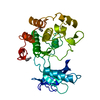
| ||||||||
|---|---|---|---|---|---|---|---|---|---|
| 1 |
| ||||||||
| Unit cell |
| ||||||||
| Atom site foot note | 1: CIS PROLINE - PRO 1071 2: RESIDUES MET 1051, MET 1076 AND ARG 1131 HAVE TWO MODELED SIDE-CHAIN CONFORMATIONS. |
-Components
| #1: Protein | Mass: 34792.805 Da / Num. of mol.: 1 Source method: isolated from a genetically manipulated source Source: (gene. exp.) Homo sapiens (human) / References: UniProt: P06213 | ||||||
|---|---|---|---|---|---|---|---|
| #2: Chemical | Ethylmercury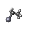  Mass: 229.651 Da / Num. of mol.: 2 / Source method: obtained synthetically / Formula: C2H5Hg Mass: 229.651 Da / Num. of mol.: 2 / Source method: obtained synthetically / Formula: C2H5Hg#3: Water | ChemComp-HOH / | Water Mass: 18.015 Da / Num. of mol.: 199 / Source method: isolated from a natural source / Formula: H2O Mass: 18.015 Da / Num. of mol.: 199 / Source method: isolated from a natural source / Formula: H2ONonpolymer details | THE MODEL INCLUDES TWO ETHYL MERCURY GROUPS (EMC) WHICH ARE COVALENTLY | Sequence details | RESIDUE NUMBERING IS ACCORDING TO EBINA ET AL. (REFERENCE 2). | |
-Experimental details
-Experiment
| Experiment | Method: X-RAY DIFFRACTION |
|---|
- Sample preparation
| Crystal | Density Matthews: 2.53 Å3/Da / Density % sol: 51.3 % | ||||||||||||||||||||
|---|---|---|---|---|---|---|---|---|---|---|---|---|---|---|---|---|---|---|---|---|---|
| Crystal grow | *PLUS Temperature: 21 ℃ / Method: vapor diffusion, hanging drop / Details: using macroseeding | ||||||||||||||||||||
| Components of the solutions | *PLUS
|
-Data collection
| Radiation | Scattering type: x-ray |
|---|---|
| Radiation wavelength | Relative weight: 1 |
| Reflection | Num. obs: 37359 / % possible obs: 93.9 % |
| Reflection | *PLUS Highest resolution: 2.1 Å / Lowest resolution: 20 Å / Observed criterion σ(I): 20.7 / Rmerge(I) obs: 0.027 |
- Processing
| Software |
| ||||||||||||||||||||||||||||||||||||||||||||||||||||||||||||||||||||||||||||||||
|---|---|---|---|---|---|---|---|---|---|---|---|---|---|---|---|---|---|---|---|---|---|---|---|---|---|---|---|---|---|---|---|---|---|---|---|---|---|---|---|---|---|---|---|---|---|---|---|---|---|---|---|---|---|---|---|---|---|---|---|---|---|---|---|---|---|---|---|---|---|---|---|---|---|---|---|---|---|---|---|---|---|
| Refinement | Resolution: 2.1→6 Å / σ(F): 2
| ||||||||||||||||||||||||||||||||||||||||||||||||||||||||||||||||||||||||||||||||
| Displacement parameters | Biso mean: 23.6 Å2 | ||||||||||||||||||||||||||||||||||||||||||||||||||||||||||||||||||||||||||||||||
| Refinement step | Cycle: LAST / Resolution: 2.1→6 Å
| ||||||||||||||||||||||||||||||||||||||||||||||||||||||||||||||||||||||||||||||||
| Refine LS restraints |
|
