 Movie
Movie Controller

[English] 日本語
 Yorodumi
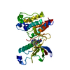
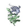
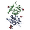
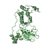
- PDB-4jv4: Crystal Structure of RIalpha(91-379) bound to HE33, a N6 di-propy... -
+ Open data

- Basic information
| Entry | Database: PDB / ID: 4jv4 | ||||||
|---|---|---|---|---|---|---|---|
| Title | Crystal Structure of RIalpha(91-379) bound to HE33, a N6 di-propyl substituted cAMP analog | ||||||
 Components Components | cAMP-dependent protein kinase type I-alpha regulatory subunit CAMP-dependent pathway CAMP-dependent pathway | ||||||
 Keywords Keywords | TRANSFERASE/TRANSFERASE INHIBITOR / cAMP-dependent protein kinase / cyclic nucleotide analogs / isoform selectivity / fluorescence anisotropy / TRANSFERASE-TRANSFERASE INHIBITOR complex | ||||||
| Function / homology |  Function and homology information Function and homology informationsperm connecting piece / PKA activation in glucagon signalling / DARPP-32 events / CREB1 phosphorylation through the activation of Adenylate Cyclase / GPER1 signaling / Factors involved in megakaryocyte development and platelet production / PKA activation / nucleotide-activated protein kinase complex / Hedgehog 'off' state / cAMP-dependent protein kinase inhibitor activity ...sperm connecting piece / PKA activation in glucagon signalling / DARPP-32 events / CREB1 phosphorylation through the activation of Adenylate Cyclase / GPER1 signaling / Factors involved in megakaryocyte development and platelet production / PKA activation / nucleotide-activated protein kinase complex / Hedgehog 'off' state / cAMP-dependent protein kinase inhibitor activity / cardiac muscle cell proliferation / cAMP-dependent protein kinase complex / sarcomere organization / Vasopressin regulates renal water homeostasis via Aquaporins / cellular response to glucagon stimulus / axoneme / negative regulation of activated T cell proliferation / protein kinase A catalytic subunit binding / plasma membrane raft / mesoderm formation / immunological synapse / cAMP binding / multivesicular body / regulation of protein phosphorylation / neuromuscular junction / positive regulation of insulin secretion / adenylate cyclase-activating G protein-coupled receptor signaling pathway / protein domain specific binding / negative regulation of gene expression / centrosome / glutamatergic synapse / ubiquitin protein ligase binding / identical protein binding / cytosol / cytoplasmSimilarity search - Function | ||||||
| Biological species |  Bos taurus (cattle) Bos taurus (cattle) | ||||||
| Method | X-RAY DIFFRACTION / SYNCHROTRON / MOLECULAR REPLACEMENT / Resolution: 2.952 Å | ||||||
 Authors Authors | Brown, S.H.J. / Cheng, C.Y. / Saldanha, A.S. / Wu, J. / Cottam, H. / Sankaran, B. / Taylor, S.S. | ||||||
 Citation Citation | Journal: Acs Chem.Biol. / Year: 2013 Title: Implementing Fluorescence Anisotropy Screening and Crystallographic Analysis to Define PKA Isoform-Selective Activation by cAMP Analogs. Authors: Brown, S.H. / Cheng, C.Y. / Saldanha, S.A. / Wu, J. / Cottam, H.B. / Sankaran, B. / Taylor, S.S. | ||||||
| History |
|
- Structure visualization
| Structure viewer | Molecule: MolmilJmol/JSmol |
|---|
- Downloads & links
-Download
| PDBx/mmCIF format | 4jv4.cif.gz | 118.2 KB | Display | PDBx/mmCIF format |
|---|---|---|---|---|
| PDB format | pdb4jv4.ent.gz | 91.5 KB | Display | PDB format |
| PDBx/mmJSON format | 4jv4.json.gz | Tree view | PDBx/mmJSON format | |
| Others |  Other downloads Other downloads |
-Validation report
| Arichive directory | https://data.pdbj.org/pub/pdb/validation_reports/jv/4jv4ftp://data.pdbj.org/pub/pdb/validation_reports/jv/4jv4 | HTTPS FTP |
|---|
-Related structure data
| Related structure data |  4jvaC  1ne6S S: Starting model for refinement C: citing same article ( |
|---|---|
| Similar structure data |


-Links
 PDBj
PDBj


- Assembly
| Deposited unit | 
| ||||||||
|---|---|---|---|---|---|---|---|---|---|
| 1 |
| ||||||||
| Unit cell |
|
-Components
| #1: Protein | CAMP-dependent pathway Mass: 32493.854 Da / Num. of mol.: 1 / Fragment: RIalpha (93-380) / Mutation: deletion mutant Source method: isolated from a genetically manipulated source Source: (gene. exp.) Bos taurus (cattle) / Gene: PRKAR1A / Plasmid: pRSET / Production host:  Escherichia coli (E. coli) / References: UniProt: P00514 Escherichia coli (E. coli) / References: UniProt: P00514 |
|---|---|
| #2: Chemical | 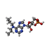  Mass: 413.365 Da / Num. of mol.: 2 / Source method: obtained synthetically / Formula: C16H24N5O6P Mass: 413.365 Da / Num. of mol.: 2 / Source method: obtained synthetically / Formula: C16H24N5O6P |
-Experimental details
-Experiment
| Experiment | Method: X-RAY DIFFRACTION / Number of used crystals: 1 |
|---|
- Sample preparation
| Crystal | Density Matthews: 3.32 Å3/Da / Density % sol: 62.91 % |
|---|---|
| Crystal grow | Temperature: 298 K / Method: vapor diffusion, hanging drop / pH: 7 Details: 6.3% PEG 3350, 0.074 M sodium malonate (pH 7.0) after 3 weeks of growth, VAPOR DIFFUSION, HANGING DROP, temperature 298.0K |
-Data collection
| Diffraction | Mean temperature: 100 K |
|---|---|
| Diffraction source | Source: SYNCHROTRON / Site: ALS  / Beamline: 8.2.1 / Wavelength: 1 Å / Beamline: 8.2.1 / Wavelength: 1 Å |
| Detector | Type: ADSC QUANTUM 315 / Detector: CCD / Date: Apr 30, 2009 |
| Radiation | Monochromator: GRAPHITE / Protocol: SINGLE WAVELENGTH / Monochromatic (M) / Laue (L): M / Scattering type: x-ray |
| Radiation wavelength | Wavelength: 1 Å / Relative weight: 1 |
| Reflection | Resolution: 2.95→50 Å / Num. obs: 9906 / % possible obs: 99.7 % / Observed criterion σ(F): 2 / Redundancy: 9.2 % / Rmerge(I) obs: 0.074 / Net I/σ(I): 40.6 |
| Reflection shell | Resolution: 2.95→3 Å / Redundancy: 7.6 % / Rmerge(I) obs: 0.417 / Mean I/σ(I) obs: 3.3 / % possible all: 99.4 |
- Processing
| Software |
| ||||||||||||||||||||||||||||||||||||||||
|---|---|---|---|---|---|---|---|---|---|---|---|---|---|---|---|---|---|---|---|---|---|---|---|---|---|---|---|---|---|---|---|---|---|---|---|---|---|---|---|---|---|
| Refinement | Method to determine structure: MOLECULAR REPLACEMENT Starting model: PDB ENTRY 1NE6 Resolution: 2.952→44.915 Å / SU ML: 0.39 / Isotropic thermal model: ANISOTROPIC / σ(F): 1.34 / Phase error: 29.15 / Stereochemistry target values: ML
| ||||||||||||||||||||||||||||||||||||||||
| Solvent computation | Shrinkage radii: 0.9 Å / VDW probe radii: 1.11 Å / Solvent model: FLAT BULK SOLVENT MODEL / Bsol: 79.532 Å2 / ksol: 0.317 e/Å3 | ||||||||||||||||||||||||||||||||||||||||
| Displacement parameters |
| ||||||||||||||||||||||||||||||||||||||||
| Refinement step | Cycle: LAST / Resolution: 2.952→44.915 Å
| ||||||||||||||||||||||||||||||||||||||||
| Refine LS restraints |
| ||||||||||||||||||||||||||||||||||||||||
| LS refinement shell |
| ||||||||||||||||||||||||||||||||||||||||
| Refinement TLS params. | Method: refined / Origin x: 6.6589 Å / Origin y: -23.0921 Å / Origin z: -0.1964 Å
| ||||||||||||||||||||||||||||||||||||||||
| Refinement TLS group | Selection details: chain A |
