 Movie
Movie Controller


+ Open data

- Basic information
| Entry | Database: PDB / ID: 4edm | ||||||
|---|---|---|---|---|---|---|---|
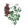
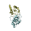
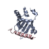
| Title | Crystal structure of beta-parvin CH2 domain | ||||||
 Components Components | Beta-parvin | ||||||
 Keywords Keywords |  SIGNALING PROTEIN / calponin homology domain / protein-protein interaction / LD motif / integrin signaling / focal adhesion / Adaptor protein / Paxillin / Integrin linked kinase SIGNALING PROTEIN / calponin homology domain / protein-protein interaction / LD motif / integrin signaling / focal adhesion / Adaptor protein / Paxillin / Integrin linked kinase | ||||||
| Function / homology |  Function and homology information Function and homology informationestablishment or maintenance of cell polarity regulating cell shape / Regulation of cytoskeletal remodeling and cell spreading by IPP complex components / Cell-extracellular matrix interactions / cell projection assembly / lamellipodium assembly / establishment or maintenance of cell polarity / substrate adhesion-dependent cell spreading / Z disc / actin cytoskeleton / lamellipodium ...establishment or maintenance of cell polarity regulating cell shape / Regulation of cytoskeletal remodeling and cell spreading by IPP complex components / Cell-extracellular matrix interactions / cell projection assembly / lamellipodium assembly / establishment or maintenance of cell polarity / substrate adhesion-dependent cell spreading / Z disc / actin cytoskeleton / lamellipodium / actin binding / actin cytoskeleton organization / focal adhesion / plasma membrane / cytosol / cytoplasmSimilarity search - Function | ||||||
| Biological species |  Homo sapiens (human) Homo sapiens (human) | ||||||
| Method | X-RAY DIFFRACTION / SYNCHROTRON / MOLECULAR REPLACEMENT / Resolution: 2 Å | ||||||
 Authors Authors | Stiegler, A.L. / Draheim, K.M. / Li, X. / Chayen, N.E. / Calderwood, D.A. / Boggon, T.J. | ||||||
 Citation Citation | Journal: J.Biol.Chem. / Year: 2012 Title: Structural basis for paxillin binding and focal adhesion targeting of beta-parvin. Authors: Stiegler, A.L. / Draheim, K.M. / Li, X. / Chayen, N.E. / Calderwood, D.A. / Boggon, T.J. | ||||||
| History |
|
- Structure visualization
| Structure viewer | Molecule: MolmilJmol/JSmol |
|---|
- Downloads & links
-Download
| PDBx/mmCIF format | 4edm.cif.gz | 120.8 KB | Display | PDBx/mmCIF format |
|---|---|---|---|---|
| PDB format | pdb4edm.ent.gz | 97.2 KB | Display | PDB format |
| PDBx/mmJSON format | 4edm.json.gz | Tree view | PDBx/mmJSON format | |
| Others |  Other downloads Other downloads |
-Validation report
| Arichive directory | https://data.pdbj.org/pub/pdb/validation_reports/ed/4edmftp://data.pdbj.org/pub/pdb/validation_reports/ed/4edm | HTTPS FTP |
|---|
-Related structure data


-Links
 PDBj
PDBj- Assembly
| Deposited unit | 
| ||||||||
|---|---|---|---|---|---|---|---|---|---|
| 1 | 
| ||||||||
| 2 | 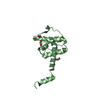
| ||||||||
| Unit cell |
|
-Components
| #1: Protein | Mass: 15371.638 Da / Num. of mol.: 2 / Fragment: C-terminal calponin homology domain Source method: isolated from a genetically manipulated source Source: (gene. exp.) Homo sapiens (human) / Gene: PARVB, CGI-56 / Plasmid: pCDFDuet-1 / Production host:  Escherichia coli (E. coli) / Strain (production host): BL21(DE3) / References: UniProt: Q9HBI1 Escherichia coli (E. coli) / Strain (production host): BL21(DE3) / References: UniProt: Q9HBI1#2: Chemical | ChemComp-EDO / Ethylene glycol  Mass: 62.068 Da / Num. of mol.: 7 / Source method: obtained synthetically / Formula: C2H6O2 Mass: 62.068 Da / Num. of mol.: 7 / Source method: obtained synthetically / Formula: C2H6O2#3: Water | ChemComp-HOH / | Water Mass: 18.015 Da / Num. of mol.: 116 / Source method: isolated from a natural source / Formula: H2O Mass: 18.015 Da / Num. of mol.: 116 / Source method: isolated from a natural source / Formula: H2O |
|---|
-Experimental details
-Experiment
| Experiment | Method: X-RAY DIFFRACTION / Number of used crystals: 1 |
|---|
- Sample preparation
| Crystal | Density Matthews: 2.86 Å3/Da / Density % sol: 57.02 % |
|---|---|
| Crystal grow | Temperature: 298 K / Method: microbatch / pH: 7.5 Details: 12% PEG 550 MME, 0.1M Tris, pH 7.5, Microbatch, temperature 298K |
-Data collection
| Diffraction | Mean temperature: 173 K |
|---|---|
| Diffraction source | Source: SYNCHROTRON / Site: NSLS  / Beamline: X6A / Wavelength: 1.0781 Å / Beamline: X6A / Wavelength: 1.0781 Å |
| Detector | Type: ADSC QUANTUM 270 / Detector: CCD / Date: Jul 2, 2010 / Details: mirror |
| Radiation | Monochromator: Si(111) channel cut monochromator / Protocol: SINGLE WAVELENGTH / Monochromatic (M) / Laue (L): M / Scattering type: x-ray |
| Radiation wavelength | Wavelength: 1.0781 Å / Relative weight: 1 |
| Reflection | Resolution: 2→50 Å / Num. obs: 23924 / % possible obs: 98.2 % / Observed criterion σ(F): 0 / Observed criterion σ(I): -3 / Redundancy: 7.8 % / Biso Wilson estimate: 40.1 Å2 / Rsym value: 0.082 / Net I/σ(I): 25.891 |
| Reflection shell | Resolution: 2→2.07 Å / Redundancy: 5.6 % / Rmerge(I) obs: 0.68 / Mean I/σ(I) obs: 1.905 / % possible all: 92.5 |
- Processing
| Software |
| |||||||||||||||||||||||||||||||||||||||||||||||||||||||||||||||||||||||||||||||||||||||||||||||||||||||||||||||||||||||||||||||||||||||||||||||||||||||||||||||||||||||||||||||||||||||||||||||||||||||||||||||||||||||||||||||||
|---|---|---|---|---|---|---|---|---|---|---|---|---|---|---|---|---|---|---|---|---|---|---|---|---|---|---|---|---|---|---|---|---|---|---|---|---|---|---|---|---|---|---|---|---|---|---|---|---|---|---|---|---|---|---|---|---|---|---|---|---|---|---|---|---|---|---|---|---|---|---|---|---|---|---|---|---|---|---|---|---|---|---|---|---|---|---|---|---|---|---|---|---|---|---|---|---|---|---|---|---|---|---|---|---|---|---|---|---|---|---|---|---|---|---|---|---|---|---|---|---|---|---|---|---|---|---|---|---|---|---|---|---|---|---|---|---|---|---|---|---|---|---|---|---|---|---|---|---|---|---|---|---|---|---|---|---|---|---|---|---|---|---|---|---|---|---|---|---|---|---|---|---|---|---|---|---|---|---|---|---|---|---|---|---|---|---|---|---|---|---|---|---|---|---|---|---|---|---|---|---|---|---|---|---|---|---|---|---|---|---|---|---|---|---|---|---|---|---|---|---|---|---|---|---|---|---|
| Refinement | Method to determine structure: MOLECULAR REPLACEMENT / Resolution: 2→50 Å / Cor.coef. Fo:Fc: 0.964 / Cor.coef. Fo:Fc free: 0.943 / Occupancy max: 1 / Occupancy min: 0.5 / SU B: 11.627 / SU ML: 0.154 / Cross valid method: THROUGHOUT / ESU R: 0.177 / ESU R Free: 0.164 / Stereochemistry target values: MAXIMUM LIKELIHOOD / Details: HYDROGENS HAVE BEEN USED IF PRESENT IN THE INPUT
| |||||||||||||||||||||||||||||||||||||||||||||||||||||||||||||||||||||||||||||||||||||||||||||||||||||||||||||||||||||||||||||||||||||||||||||||||||||||||||||||||||||||||||||||||||||||||||||||||||||||||||||||||||||||||||||||||
| Solvent computation | Ion probe radii: 0.8 Å / Shrinkage radii: 0.8 Å / VDW probe radii: 1.2 Å / Solvent model: MASK | |||||||||||||||||||||||||||||||||||||||||||||||||||||||||||||||||||||||||||||||||||||||||||||||||||||||||||||||||||||||||||||||||||||||||||||||||||||||||||||||||||||||||||||||||||||||||||||||||||||||||||||||||||||||||||||||||
| Displacement parameters | Biso max: 129.56 Å2 / Biso mean: 58.1985 Å2 / Biso min: 25.27 Å2
| |||||||||||||||||||||||||||||||||||||||||||||||||||||||||||||||||||||||||||||||||||||||||||||||||||||||||||||||||||||||||||||||||||||||||||||||||||||||||||||||||||||||||||||||||||||||||||||||||||||||||||||||||||||||||||||||||
| Refinement step | Cycle: LAST / Resolution: 2→50 Å
| |||||||||||||||||||||||||||||||||||||||||||||||||||||||||||||||||||||||||||||||||||||||||||||||||||||||||||||||||||||||||||||||||||||||||||||||||||||||||||||||||||||||||||||||||||||||||||||||||||||||||||||||||||||||||||||||||
| Refine LS restraints |
| |||||||||||||||||||||||||||||||||||||||||||||||||||||||||||||||||||||||||||||||||||||||||||||||||||||||||||||||||||||||||||||||||||||||||||||||||||||||||||||||||||||||||||||||||||||||||||||||||||||||||||||||||||||||||||||||||
| LS refinement shell | Resolution: 2→2.054 Å / Total num. of bins used: 20
| |||||||||||||||||||||||||||||||||||||||||||||||||||||||||||||||||||||||||||||||||||||||||||||||||||||||||||||||||||||||||||||||||||||||||||||||||||||||||||||||||||||||||||||||||||||||||||||||||||||||||||||||||||||||||||||||||
| Refinement TLS params. | Method: refined / Refine-ID: X-RAY DIFFRACTION
| |||||||||||||||||||||||||||||||||||||||||||||||||||||||||||||||||||||||||||||||||||||||||||||||||||||||||||||||||||||||||||||||||||||||||||||||||||||||||||||||||||||||||||||||||||||||||||||||||||||||||||||||||||||||||||||||||
| Refinement TLS group |
|
