 Movie
Movie Controller


+ Open data

- Basic information
| Entry | Database: PDB / ID: 6pir | ||||||
|---|---|---|---|---|---|---|---|
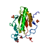
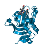
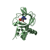
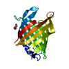
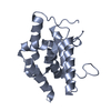
| Title | Crystal structure of the Legionella effector protein MavE | ||||||
 Components Components | MavE | ||||||
 Keywords Keywords |  SIGNALING PROTEIN / Twinned two-helix bundle / NPxY motif / phosphotyrosine binding / membrane-anchored SIGNALING PROTEIN / Twinned two-helix bundle / NPxY motif / phosphotyrosine binding / membrane-anchored | ||||||
| Function / homology | membrane => GO:0016020 / CITRIC ACID / Uncharacterized protein Function and homology information Function and homology information | ||||||
| Biological species |  Legionella pneumophila subsp. pneumophila (bacteria) Legionella pneumophila subsp. pneumophila (bacteria) | ||||||
| Method | X-RAY DIFFRACTION / SYNCHROTRON / SAD / Resolution: 1.65 Å | ||||||
 Authors Authors | Cygler, M. / Voth, K.A. | ||||||
| Funding support |  Canada, 1items Canada, 1items
| ||||||
 Citation Citation | Journal: To Be Published Title: Crystal structure of the Legionella effector MavE Authors: Cygler, M. / Voth, K.A. | ||||||
| History |
|
- Structure visualization
| Structure viewer | Molecule: MolmilJmol/JSmol |
|---|
- Downloads & links
-Download
| PDBx/mmCIF format | 6pir.cif.gz | 176.5 KB | Display | PDBx/mmCIF format |
|---|---|---|---|---|
| PDB format | pdb6pir.ent.gz | 140.9 KB | Display | PDB format |
| PDBx/mmJSON format | 6pir.json.gz | Tree view | PDBx/mmJSON format | |
| Others |  Other downloads Other downloads |
-Validation report
| Arichive directory | https://data.pdbj.org/pub/pdb/validation_reports/pi/6pirftp://data.pdbj.org/pub/pdb/validation_reports/pi/6pir | HTTPS FTP |
|---|
-Related structure data
| Similar structure data |
|---|


-Links
 PDBj
PDBj- Assembly
| Deposited unit | 
| ||||||||
|---|---|---|---|---|---|---|---|---|---|
| 1 | 
| ||||||||
| 2 | 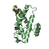
| ||||||||
| 3 | 
| ||||||||
| 4 | 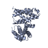
| ||||||||
| 5 | 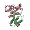
| ||||||||
| Unit cell |
| ||||||||
| Components on special symmetry positions |
|
-Components
| #1: Protein | Mass: 15839.636 Da / Num. of mol.: 3 Source method: isolated from a genetically manipulated source Source: (gene. exp.) Legionella pneumophila subsp. pneumophila (strain Philadelphia 1 / ATCC 33152 / DSM 7513) (bacteria)Strain: Philadelphia 1 / ATCC 33152 / DSM 7513 / Gene: lpg2344 / Production host: Escherichia coli BL21(DE3) (bacteria) / Strain (production host): BL21(DE3) / References: UniProt: Q5ZT21#2: Chemical | Citric acid  Mass: 192.124 Da / Num. of mol.: 2 / Source method: obtained synthetically / Formula: C6H8O7 Mass: 192.124 Da / Num. of mol.: 2 / Source method: obtained synthetically / Formula: C6H8O7#3: Water | ChemComp-HOH / | Water Mass: 18.015 Da / Num. of mol.: 252 / Source method: isolated from a natural source / Formula: H2O Mass: 18.015 Da / Num. of mol.: 252 / Source method: isolated from a natural source / Formula: H2OHas ligand of interest | N | |
|---|
-Experimental details
-Experiment
| Experiment | Method: X-RAY DIFFRACTION / Number of used crystals: 1 |
|---|
- Sample preparation
| Crystal | Density Matthews: 2.04 Å3/Da / Density % sol: 39.78 % |
|---|---|
| Crystal grow | Temperature: 293.15 K / Method: vapor diffusion, hanging drop / Details: 0.1M Citrate, pH 6.0, 18% PEG20K / PH range: 6 |
-Data collection
| Diffraction | Mean temperature: 293.15 K / Serial crystal experiment: N |
|---|---|
| Diffraction source | Source: SYNCHROTRON / Site: CLSI / Beamline: 08ID-1 / Wavelength: 1 Å |
| Detector | Type: DECTRIS PILATUS3 S 6M / Detector: PIXEL / Date: Nov 26, 2016 |
| Radiation | Protocol: SINGLE WAVELENGTH / Monochromatic (M) / Laue (L): M / Scattering type: x-ray |
| Radiation wavelength | Wavelength: 1 Å / Relative weight: 1 |
| Reflection | Resolution: 1.65→48.77 Å / Num. obs: 82310 / % possible obs: 99.3 % / Redundancy: 6.96 % / CC1/2: 0.999 / Net I/σ(I): 15.19 |
| Reflection shell | Resolution: 1.65→1.69 Å / Num. unique obs: 13055 / CC1/2: 0.593 |
- Processing
| Software |
| |||||||||||||||||||||||||||||||||||||||||||||||||||||||||||||||||||||||||||||||||||||||||||||||||||||||||
|---|---|---|---|---|---|---|---|---|---|---|---|---|---|---|---|---|---|---|---|---|---|---|---|---|---|---|---|---|---|---|---|---|---|---|---|---|---|---|---|---|---|---|---|---|---|---|---|---|---|---|---|---|---|---|---|---|---|---|---|---|---|---|---|---|---|---|---|---|---|---|---|---|---|---|---|---|---|---|---|---|---|---|---|---|---|---|---|---|---|---|---|---|---|---|---|---|---|---|---|---|---|---|---|---|---|---|
| Refinement | Method to determine structure: SAD / Resolution: 1.65→46.267 Å / SU ML: 0.23 / Cross valid method: FREE R-VALUE / σ(F): 1.36 / Phase error: 28.41
| |||||||||||||||||||||||||||||||||||||||||||||||||||||||||||||||||||||||||||||||||||||||||||||||||||||||||
| Solvent computation | Shrinkage radii: 0.9 Å / VDW probe radii: 1.11 Å | |||||||||||||||||||||||||||||||||||||||||||||||||||||||||||||||||||||||||||||||||||||||||||||||||||||||||
| Refinement step | Cycle: LAST / Resolution: 1.65→46.267 Å
| |||||||||||||||||||||||||||||||||||||||||||||||||||||||||||||||||||||||||||||||||||||||||||||||||||||||||
| Refine LS restraints |
| |||||||||||||||||||||||||||||||||||||||||||||||||||||||||||||||||||||||||||||||||||||||||||||||||||||||||
| LS refinement shell |
| |||||||||||||||||||||||||||||||||||||||||||||||||||||||||||||||||||||||||||||||||||||||||||||||||||||||||
| Refinement TLS params. | Method: refined / Refine-ID: X-RAY DIFFRACTION
| |||||||||||||||||||||||||||||||||||||||||||||||||||||||||||||||||||||||||||||||||||||||||||||||||||||||||
| Refinement TLS group |
|
