 Movie
Movie Controller

[English] 日本語
 Yorodumi
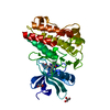
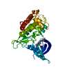
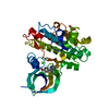
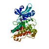
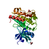
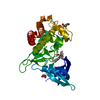
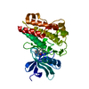
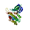
- PDB-5qil: TGF-BETA RECEPTOR TYPE 1 KINASE DOMAIN (T204D) IN COMPLEX WITH N-... -
+ Open data

- Basic information
| Entry | Database: PDB / ID: 5qil | ||||||
|---|---|---|---|---|---|---|---|
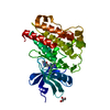
| Title | TGF-BETA RECEPTOR TYPE 1 KINASE DOMAIN (T204D) IN COMPLEX WITH N-{4-[3-(6-METHOXYPYRIDIN-3-YL)-1H-PYRROLO[3,2-B]PYRIDIN-2-YL]PYRIDIN-2-YL}ACETAMIDE | ||||||
 Components Components | TGF-beta receptor type-1 | ||||||
 Keywords Keywords | TRANSFERASE/TRANSFERASE INHIBITOR /  ALK5 / SB431542 / KINASE DOMAIN / TRANSFERASE-TRANSFERASE INHIBITOR COMPLEX / TRANSFERASE ALK5 / SB431542 / KINASE DOMAIN / TRANSFERASE-TRANSFERASE INHIBITOR COMPLEX / TRANSFERASE | ||||||
| Function / homology |  Function and homology information Function and homology informationextracellular structure organization / epicardium morphogenesis / parathyroid gland development / transforming growth factor beta ligand-receptor complex / regulation of cardiac muscle cell proliferation / myofibroblast differentiation / positive regulation of tight junction disassembly / positive regulation of epithelial to mesenchymal transition involved in endocardial cushion formation / TGFBR2 Kinase Domain Mutants in Cancer / transforming growth factor beta receptor activity ...extracellular structure organization / epicardium morphogenesis / parathyroid gland development / transforming growth factor beta ligand-receptor complex / regulation of cardiac muscle cell proliferation / myofibroblast differentiation / positive regulation of tight junction disassembly / positive regulation of epithelial to mesenchymal transition involved in endocardial cushion formation / TGFBR2 Kinase Domain Mutants in Cancer / transforming growth factor beta receptor activity / positive regulation of mesenchymal stem cell proliferation / ventricular compact myocardium morphogenesis / SMAD2/3 Phosphorylation Motif Mutants in Cancer / TGFBR1 KD Mutants in Cancer / positive regulation of vasculature development / regulation of epithelial to mesenchymal transition / activin receptor activity, type I / cardiac epithelial to mesenchymal transition / transforming growth factor beta receptor activity, type I / activin receptor complex / mesenchymal cell differentiation / neuron fate commitment / positive regulation of extracellular matrix assembly / type II transforming growth factor beta receptor binding / TGFBR1 LBD Mutants in Cancer / angiogenesis involved in coronary vascular morphogenesis / receptor protein serine/threonine kinase / germ cell migration / transmembrane receptor protein serine/threonine kinase activity / pharyngeal system development / activin binding / coronary artery morphogenesis / activin receptor signaling pathway / ventricular trabecula myocardium morphogenesis / filopodium assembly / transforming growth factor beta binding / embryonic cranial skeleton morphogenesis / response to cholesterol / I-SMAD binding / collagen fibril organization / negative regulation of chondrocyte differentiation / endothelial cell activation / skeletal system morphogenesis / lens development in camera-type eye / endothelial cell proliferation / positive regulation of filopodium assembly / anterior/posterior pattern specification / artery morphogenesis / ventricular septum morphogenesis / roof of mouth development / SMAD binding / TGF-beta receptor signaling activates SMADs / negative regulation of endothelial cell proliferation / positive regulation of SMAD protein signal transduction / blastocyst development / regulation of protein ubiquitination / endothelial cell migration / bicellular tight junction / epithelial to mesenchymal transition / positive regulation of epithelial to mesenchymal transition / positive regulation of stress fiber assembly / cellular response to transforming growth factor beta stimulus / positive regulation of endothelial cell proliferation / Downregulation of TGF-beta receptor signaling / negative regulation of cell migration / transforming growth factor beta receptor signaling pathway / post-embryonic development / thymus development / TGF-beta receptor signaling in EMT (epithelial to mesenchymal transition) / skeletal system development / kidney development / positive regulation of apoptotic signaling pathway / cell motility / negative regulation of extrinsic apoptotic signaling pathway / wound healing / cellular response to growth factor stimulus / UCH proteinases / male gonad development / nervous system development / heart development / positive regulation of cell growth / regulation of gene expression / peptidyl-serine phosphorylation / in utero embryonic development / positive regulation of phosphatidylinositol 3-kinase/protein kinase B signal transduction / receptor complex / Ub-specific processing proteases / regulation of cell cycle / endosome / protein kinase activity / intracellular signal transduction / positive regulation of cell migration / membrane raft / protein serine/threonine kinase activity / apoptotic process / ubiquitin protein ligase binding / positive regulation of cell population proliferation / regulation of DNA-templated transcription / positive regulation of gene expression / positive regulation of DNA-templated transcriptionSimilarity search - Function | ||||||
| Biological species |  Homo sapiens (human) Homo sapiens (human) | ||||||
| Method | X-RAY DIFFRACTION / SYNCHROTRON / MOLECULAR REPLACEMENT / molecular replacement / Resolution: 1.98 Å | ||||||
 Authors Authors | Sheriff, S. | ||||||
 Citation Citation | Journal: ACS Med Chem Lett / Year: 2018 Title: Discovery of 4-Azaindole Inhibitors of TGF beta RI as Immuno-oncology Agents. Authors: Zhang, Y. / Zhao, Y. / Tebben, A.J. / Sheriff, S. / Ruzanov, M. / Fereshteh, M.P. / Fan, Y. / Lippy, J. / Swanson, J. / Ho, C.P. / Wautlet, B.S. / Rose, A. / Parrish, K. / Yang, Z. / ...Authors: Zhang, Y. / Zhao, Y. / Tebben, A.J. / Sheriff, S. / Ruzanov, M. / Fereshteh, M.P. / Fan, Y. / Lippy, J. / Swanson, J. / Ho, C.P. / Wautlet, B.S. / Rose, A. / Parrish, K. / Yang, Z. / Donnell, A.F. / Zhang, L. / Fink, B.E. / Vite, G.D. / Augustine-Rauch, K. / Fargnoli, J. / Borzilleri, R.M. | ||||||
| History |
|
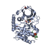
- Structure visualization
| Structure viewer | Molecule: MolmilJmol/JSmol |
|---|
- Downloads & links
-Download
| PDBx/mmCIF format | 5qil.cif.gz | 78.6 KB | Display | PDBx/mmCIF format |
|---|---|---|---|---|
| PDB format | pdb5qil.ent.gz | 57.5 KB | Display | PDB format |
| PDBx/mmJSON format | 5qil.json.gz | Tree view | PDBx/mmJSON format | |
| Others |  Other downloads Other downloads |
-Validation report
| Arichive directory | https://data.pdbj.org/pub/pdb/validation_reports/qi/5qilftp://data.pdbj.org/pub/pdb/validation_reports/qi/5qil | HTTPS FTP |
|---|
-Group deposition
| ID | G_1002052 (4 entries) |
|---|---|
| Title | TGFBR |
| Type | undefined |
| Description | Structures deposited in support of Y. Zhang et al., ACS Med.Chem.Lett., about to be submitted |

-Related structure data
| Related structure data |  3tzmS S: Starting model for refinement |
|---|---|
| Similar structure data |


-Links
 PDBj
PDBj


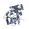
- Assembly
| Deposited unit | 
| ||||||||
|---|---|---|---|---|---|---|---|---|---|
| 1 |
| ||||||||
| Unit cell |
|
-Components
| #1: Protein | Mass: 35165.473 Da / Num. of mol.: 1 / Fragment: KINASE DOMAIN, UNP RESIDUES 200-503 / Mutation: T204D Source method: isolated from a genetically manipulated source Source: (gene. exp.) Homo sapiens (human) / Gene: TGFBR1, ALK5, SKR4 / Plasmid: PFASTBAC1 / Production host:   SPODOPTERA FRUGIPERDA (fall armyworm) SPODOPTERA FRUGIPERDA (fall armyworm)References: UniProt: P36897, receptor protein serine/threonine kinase |
|---|---|
| #2: Chemical | ChemComp-J2V /   Mass: 359.381 Da / Num. of mol.: 1 / Source method: obtained synthetically / Formula: C20H17N5O2 Mass: 359.381 Da / Num. of mol.: 1 / Source method: obtained synthetically / Formula: C20H17N5O2 |
| #3: Water | ChemComp-HOH / Water Mass: 18.015 Da / Num. of mol.: 156 / Source method: isolated from a natural source / Formula: H2O Mass: 18.015 Da / Num. of mol.: 156 / Source method: isolated from a natural source / Formula: H2O |
-Experimental details
-Experiment
| Experiment | Method: X-RAY DIFFRACTION / Number of used crystals: 1 |
|---|
- Sample preparation
| Crystal | Density Matthews: 2.03 Å3/Da / Density % sol: 39.35 % |
|---|---|
| Crystal grow | Temperature: 277 K / Method: vapor diffusion, hanging drop / pH: 5.6 / Details: 23%(w/v) PEG3350, 3%(v/v) glcyerol |
-Data collection
| Diffraction | Mean temperature: 100 K |
|---|---|
| Diffraction source | Source: SYNCHROTRON / Site: APS  / Beamline: 17-ID / Wavelength: 1 / Wavelength: 1 Å / Beamline: 17-ID / Wavelength: 1 / Wavelength: 1 Å |
| Detector | Type: DECTRIS PILATUS 6M / Detector: PIXEL / Date: Feb 19, 2015 |
| Radiation | Protocol: SINGLE WAVELENGTH / Monochromatic (M) / Laue (L): M / Scattering type: x-ray |
| Radiation wavelength | Wavelength: 1 Å / Relative weight: 1 |
| Reflection | Resolution: 1.98→41.6 Å / Num. obs: 20725 / % possible obs: 99.9 % / Observed criterion σ(I): 0 / Redundancy: 6.4 % / Biso Wilson estimate: 24.27 Å2 / Rsym value: 0.092 / Net I/σ(I): 14.5 |
| Reflection shell | Resolution: 1.98→2.29 Å / Redundancy: 6.5 % / Mean I/σ(I) obs: 5.5 / Rsym value: 0.365 / Rejects: 0 / % possible all: 100 |
-Phasing
| Phasing | Method: molecular replacement |
|---|
- Processing
| Software |
| ||||||||||||||||||||||||||||||||||||||||||||||||||||||||||||||||||||||||||||||||||||||||||||||||||||||||||||
|---|---|---|---|---|---|---|---|---|---|---|---|---|---|---|---|---|---|---|---|---|---|---|---|---|---|---|---|---|---|---|---|---|---|---|---|---|---|---|---|---|---|---|---|---|---|---|---|---|---|---|---|---|---|---|---|---|---|---|---|---|---|---|---|---|---|---|---|---|---|---|---|---|---|---|---|---|---|---|---|---|---|---|---|---|---|---|---|---|---|---|---|---|---|---|---|---|---|---|---|---|---|---|---|---|---|---|---|---|---|
| Refinement | Method to determine structure: MOLECULAR REPLACEMENT Starting model: 3TZM Resolution: 1.98→20.8 Å / Cor.coef. Fo:Fc: 0.9251 / Cor.coef. Fo:Fc free: 0.9178 / SU R Cruickshank DPI: 0.17 / Cross valid method: THROUGHOUT / σ(F): 0 / SU R Blow DPI: 0.179 / SU Rfree Blow DPI: 0.15 / SU Rfree Cruickshank DPI: 0.147
| ||||||||||||||||||||||||||||||||||||||||||||||||||||||||||||||||||||||||||||||||||||||||||||||||||||||||||||
| Displacement parameters | Biso max: 110.35 Å2 / Biso mean: 27.44 Å2 / Biso min: 9.34 Å2
| ||||||||||||||||||||||||||||||||||||||||||||||||||||||||||||||||||||||||||||||||||||||||||||||||||||||||||||
| Refine analyze | Luzzati coordinate error obs: 0.188 Å | ||||||||||||||||||||||||||||||||||||||||||||||||||||||||||||||||||||||||||||||||||||||||||||||||||||||||||||
| Refinement step | Cycle: final / Resolution: 1.98→20.8 Å
| ||||||||||||||||||||||||||||||||||||||||||||||||||||||||||||||||||||||||||||||||||||||||||||||||||||||||||||
| Refine LS restraints |
| ||||||||||||||||||||||||||||||||||||||||||||||||||||||||||||||||||||||||||||||||||||||||||||||||||||||||||||
| LS refinement shell | Resolution: 1.98→2.09 Å / Total num. of bins used: 10
|
