 Movie
Movie Controller


+ Open data

- Basic information
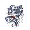
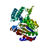
| Entry | Database: PDB / ID: 1gzo | ||||||
|---|---|---|---|---|---|---|---|
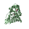
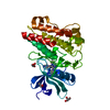
| Title | Structure of protein kinase B unphosphorylated | ||||||
 Components Components | RAC-BETA SERINE/THREONINE PROTEIN KINASE | ||||||
 Keywords Keywords |  KINASE / TRANSFERASE / SERINE/THREONINE-PROTEIN KINASE / ATP-BINDING KINASE / TRANSFERASE / SERINE/THREONINE-PROTEIN KINASE / ATP-BINDING | ||||||
| Function / homology |  Function and homology information Function and homology informationretinal rod cell apoptotic process / PDE3B signalling / cellular response to high light intensity / Inhibition of TSC complex formation by PKB / AKT-mediated inactivation of FOXO1A / Negative regulation of the PI3K/AKT network / negative regulation of long-chain fatty acid import across plasma membrane / Activation of AKT2 / AKT phosphorylates targets in the nucleus / : ...retinal rod cell apoptotic process / PDE3B signalling / cellular response to high light intensity / Inhibition of TSC complex formation by PKB / AKT-mediated inactivation of FOXO1A / Negative regulation of the PI3K/AKT network / negative regulation of long-chain fatty acid import across plasma membrane / Activation of AKT2 / AKT phosphorylates targets in the nucleus / : / RUNX2 regulates genes involved in cell migration / positive regulation of fatty acid beta-oxidation / mammary gland epithelial cell differentiation / RAB GEFs exchange GTP for GDP on RABs / positive regulation of glucose metabolic process / peripheral nervous system myelin maintenance / glycogen biosynthetic process / positive regulation of cell motility / AKT phosphorylates targets in the cytosol / Regulation of TP53 Activity through Association with Co-factors / CTLA4 inhibitory signaling / fat cell differentiation / Constitutive Signaling by AKT1 E17K in Cancer / CD28 dependent PI3K/Akt signaling / Regulation of localization of FOXO transcription factors / Estrogen-dependent nuclear events downstream of ESR-membrane signaling / positive regulation of glycogen biosynthetic process / Activation of BAD and translocation to mitochondria / positive regulation of protein targeting to membrane / SARS-CoV-2 targets host intracellular signalling and regulatory pathways / Cyclin E associated events during G1/S transition / Cyclin A:Cdk2-associated events at S phase entry / regulation of cell migration / Regulation of TP53 Activity through Acetylation / FLT3 Signaling / Downregulation of ERBB2:ERBB3 signaling / VEGFR2 mediated vascular permeability / molecular function activator activity / protein localization to plasma membrane / Translocation of SLC2A4 (GLUT4) to the plasma membrane / Deactivation of the beta-catenin transactivating complex / positive regulation of glucose import / TP53 Regulates Metabolic Genes / protein modification process / ruffle membrane / Regulation of PTEN stability and activity / G beta:gamma signalling through PI3Kgamma / cellular response to insulin stimulus / glucose metabolic process / KEAP1-NFE2L2 pathway / Regulation of TP53 Degradation / PIP3 activates AKT signaling / insulin receptor signaling pathway / regulation of translation / cell cortex / early endosome / non-specific serine/threonine protein kinase / regulation of cell cycle / intracellular signal transduction / positive regulation of cell migration / phosphorylation / intracellular membrane-bounded organelle / protein serine kinase activity / protein serine/threonine kinase activity / signal transduction / nucleoplasm / ATP binding / metal ion binding / nucleus / plasma membrane / cytosolSimilarity search - Function | ||||||
| Biological species |  HOMO SAPIENS (human) HOMO SAPIENS (human) | ||||||
| Method | X-RAY DIFFRACTION / SYNCHROTRON / MOLECULAR REPLACEMENT / Resolution: 2.75 Å | ||||||
 Authors Authors | Barford, D. / Yang, J. / Hemmings, B.A. | ||||||
 Citation Citation | Journal: Mol.Cell / Year: 2002 Title: Molecular Mechanism for the Regulation of Protein Kinase B/Akt by Hydrophobic Motif Phosphorylation Authors: Yang, J. / Cron, P. / Thompson, V. / Good, V. / Hess, D. / Hemmings, B.A. / Barford, D. | ||||||
| History |
|
- Structure visualization
| Structure viewer | Molecule: MolmilJmol/JSmol |
|---|
- Downloads & links
-Download
| PDBx/mmCIF format | 1gzo.cif.gz | 71.8 KB | Display | PDBx/mmCIF format |
|---|---|---|---|---|
| PDB format | pdb1gzo.ent.gz | 52.2 KB | Display | PDB format |
| PDBx/mmJSON format | 1gzo.json.gz | Tree view | PDBx/mmJSON format | |
| Others |  Other downloads Other downloads |
-Validation report
| Arichive directory | https://data.pdbj.org/pub/pdb/validation_reports/gz/1gzoftp://data.pdbj.org/pub/pdb/validation_reports/gz/1gzo | HTTPS FTP |
|---|
-Related structure data
| Related structure data | 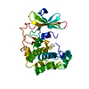 1gzkC 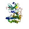 1gznC  1cdkS C: citing same article ( S: Starting model for refinement |
|---|---|
| Similar structure data |


-Links
 PDBj
PDBj


- Assembly
| Deposited unit | 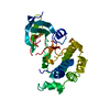
| ||||||||
|---|---|---|---|---|---|---|---|---|---|
| 1 |
| ||||||||
| Unit cell |
|
-Components
| #1: Protein | Mass: 36490.875 Da / Num. of mol.: 1 Fragment: KINASE DOMAIN WITHOUT HYDROPHOBIC MOTIF, RESIDUES 146-460 Source method: isolated from a genetically manipulated source Source: (gene. exp.) HOMO SAPIENS (human) / Production host:   SPODOPTERA FRUGIPERDA (fall armyworm) SPODOPTERA FRUGIPERDA (fall armyworm)References: UniProt: P31751, Transferases; Transferring phosphorus-containing groups; Phosphotransferases with an alcohol group as acceptor |
|---|---|
| #2: Water | ChemComp-HOH / Water Mass: 18.015 Da / Num. of mol.: 95 / Source method: isolated from a natural source / Formula: H2O Mass: 18.015 Da / Num. of mol.: 95 / Source method: isolated from a natural source / Formula: H2O |
| Compound details | FUNCTION: PROTEIN KINASE PHOSPHORYLATING SEVERAL KNOWN PROTEINS. TISSUE SPECIFICITY: ALL HUMAN CELL ...FUNCTION: PROTEIN KINASE PHOSPHORYL |
-Experimental details
-Experiment
| Experiment | Method: X-RAY DIFFRACTION / Number of used crystals: 1 |
|---|
- Sample preparation
| Crystal | Density Matthews: 2.6 Å3/Da / Density % sol: 47 % | ||||||||||||||||||||||||||||||||||||||||||
|---|---|---|---|---|---|---|---|---|---|---|---|---|---|---|---|---|---|---|---|---|---|---|---|---|---|---|---|---|---|---|---|---|---|---|---|---|---|---|---|---|---|---|---|
| Crystal grow | pH: 7.5 Details: 30% PEG 8000, 0.2 M LITHIUM SULPHATE, 0.1 M TRIS, 10MG/ML PROTEIN, pH 7.50 | ||||||||||||||||||||||||||||||||||||||||||
| Crystal grow | *PLUS Temperature: 20 ℃ / pH: 8.5 / Method: batch method | ||||||||||||||||||||||||||||||||||||||||||
| Components of the solutions | *PLUS
|
-Data collection
| Diffraction | Mean temperature: 100 K |
|---|---|
| Diffraction source | Source: SYNCHROTRON / Site: ESRF  / Beamline: ID14-4 / Wavelength: 0.92 / Beamline: ID14-4 / Wavelength: 0.92 |
| Detector | Type: ADSC CCD / Detector: CCD / Date: Sep 15, 2001 |
| Radiation | Protocol: SINGLE WAVELENGTH / Monochromatic (M) / Laue (L): M / Scattering type: x-ray |
| Radiation wavelength | Wavelength: 0.92 Å / Relative weight: 1 |
| Reflection | Resolution: 2.75→30 Å / Num. obs: 12147 / % possible obs: 94.2 % / Observed criterion σ(I): 0 / Redundancy: 4.2 % / Biso Wilson estimate: 104.6 Å2 / Rmerge(I) obs: 0.062 / Net I/σ(I): 18 |
| Reflection shell | Resolution: 2.75→2.85 Å / % possible all: 84 |
| Reflection | *PLUS Highest resolution: 2.7 Å / Lowest resolution: 30 Å / Num. measured all: 50875 / Rmerge(I) obs: 0.065 |
| Reflection shell | *PLUS % possible obs: 84 % / Rmerge(I) obs: 0.236 |
- Processing
| Software |
| ||||||||||||||||||||||||||||||||||||||||||||||||||||||||||||||||||||||||||||||||
|---|---|---|---|---|---|---|---|---|---|---|---|---|---|---|---|---|---|---|---|---|---|---|---|---|---|---|---|---|---|---|---|---|---|---|---|---|---|---|---|---|---|---|---|---|---|---|---|---|---|---|---|---|---|---|---|---|---|---|---|---|---|---|---|---|---|---|---|---|---|---|---|---|---|---|---|---|---|---|---|---|---|
| Refinement | Method to determine structure: MOLECULAR REPLACEMENT Starting model: PDB ENTRY 1CDK Resolution: 2.75→25.67 Å / Rfactor Rfree error: 0.009 / Data cutoff high absF: 1488479.19 / Isotropic thermal model: RESTRAINED / Cross valid method: THROUGHOUT / σ(F): 0
| ||||||||||||||||||||||||||||||||||||||||||||||||||||||||||||||||||||||||||||||||
| Solvent computation | Solvent model: FLAT MODEL / Bsol: 45.6653 Å2 / ksol: 0.308053 e/Å3 | ||||||||||||||||||||||||||||||||||||||||||||||||||||||||||||||||||||||||||||||||
| Displacement parameters | Biso mean: 57.7 Å2
| ||||||||||||||||||||||||||||||||||||||||||||||||||||||||||||||||||||||||||||||||
| Refine analyze |
| ||||||||||||||||||||||||||||||||||||||||||||||||||||||||||||||||||||||||||||||||
| Refinement step | Cycle: LAST / Resolution: 2.75→25.67 Å
| ||||||||||||||||||||||||||||||||||||||||||||||||||||||||||||||||||||||||||||||||
| Refine LS restraints |
| ||||||||||||||||||||||||||||||||||||||||||||||||||||||||||||||||||||||||||||||||
| LS refinement shell | Resolution: 2.75→2.92 Å / Rfactor Rfree error: 0.033 / Total num. of bins used: 6
| ||||||||||||||||||||||||||||||||||||||||||||||||||||||||||||||||||||||||||||||||
| Refinement | *PLUS Lowest resolution: 35 Å / % reflection Rfree: 9.9 % / Rfactor Rfree: 0.3 / Rfactor Rwork: 0.238 | ||||||||||||||||||||||||||||||||||||||||||||||||||||||||||||||||||||||||||||||||
| Solvent computation | *PLUS | ||||||||||||||||||||||||||||||||||||||||||||||||||||||||||||||||||||||||||||||||
| Displacement parameters | *PLUS | ||||||||||||||||||||||||||||||||||||||||||||||||||||||||||||||||||||||||||||||||
| Refine LS restraints | *PLUS
|
