 Movie
Movie Controller

[English] 日本語
 Yorodumi
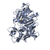
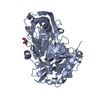
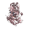
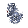
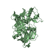
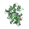
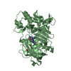
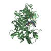
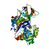
- PDB-4h3f: Structure of BACE Bound to 3-(5-((7aR)-2-imino-6-(6-methoxypyridi... -
+ Open data

- Basic information
| Entry | Database: PDB / ID: 4h3f | ||||||
|---|---|---|---|---|---|---|---|
| Title | Structure of BACE Bound to 3-(5-((7aR)-2-imino-6-(6-methoxypyridin-2-yl)-3-methyl-4-oxooctahydro-1H-pyrrolo[3,4-d]pyrimidin-7a-yl)thiophen-3-yl)benzonitrile | ||||||
 Components Components | Beta-secretase 1 | ||||||
 Keywords Keywords | HYDROLASE/HYDROLASE INHIBITOR / BACE1 / Alzheimers / HYDROLASE-HYDROLASE INHIBITOR complex | ||||||
| Function / homology |  Function and homology informationmemapsin 2 / Golgi-associated vesicle lumen / signaling receptor ligand precursor processing / beta-aspartyl-peptidase activity / amyloid precursor protein catabolic process / amyloid-beta formation / membrane protein ectodomain proteolysis / cellular response to manganese ion / prepulse inhibition / detection of mechanical stimulus involved in sensory perception of pain ...memapsin 2 / Golgi-associated vesicle lumen / signaling receptor ligand precursor processing / beta-aspartyl-peptidase activity / amyloid precursor protein catabolic process / amyloid-beta formation / membrane protein ectodomain proteolysis / cellular response to manganese ion / prepulse inhibition / detection of mechanical stimulus involved in sensory perception of pain / amyloid-beta metabolic process / cellular response to copper ion / hippocampal mossy fiber to CA3 synapse / presynaptic modulation of chemical synaptic transmission / multivesicular body / response to lead ion / trans-Golgi network / protein processing / recycling endosome / cellular response to amyloid-beta / positive regulation of neuron apoptotic process / synaptic vesicle / late endosome / amyloid-beta binding / peptidase activity / endopeptidase activity / amyloid fibril formation / lysosome / aspartic-type endopeptidase activity / early endosome / endosome membrane / endosome / membrane raft / Amyloid fiber formation / axon / endoplasmic reticulum lumen / neuronal cell body / dendrite / Golgi apparatus / enzyme binding / cell surface / proteolysis / membrane / plasma membrane Function and homology informationmemapsin 2 / Golgi-associated vesicle lumen / signaling receptor ligand precursor processing / beta-aspartyl-peptidase activity / amyloid precursor protein catabolic process / amyloid-beta formation / membrane protein ectodomain proteolysis / cellular response to manganese ion / prepulse inhibition / detection of mechanical stimulus involved in sensory perception of pain ...memapsin 2 / Golgi-associated vesicle lumen / signaling receptor ligand precursor processing / beta-aspartyl-peptidase activity / amyloid precursor protein catabolic process / amyloid-beta formation / membrane protein ectodomain proteolysis / cellular response to manganese ion / prepulse inhibition / detection of mechanical stimulus involved in sensory perception of pain / amyloid-beta metabolic process / cellular response to copper ion / hippocampal mossy fiber to CA3 synapse / presynaptic modulation of chemical synaptic transmission / multivesicular body / response to lead ion / trans-Golgi network / protein processing / recycling endosome / cellular response to amyloid-beta / positive regulation of neuron apoptotic process / synaptic vesicle / late endosome / amyloid-beta binding / peptidase activity / endopeptidase activity / amyloid fibril formation / lysosome / aspartic-type endopeptidase activity / early endosome / endosome membrane / endosome / membrane raft / Amyloid fiber formation / axon / endoplasmic reticulum lumen / neuronal cell body / dendrite / Golgi apparatus / enzyme binding / cell surface / proteolysis / membrane / plasma membraneSimilarity search - Function | ||||||
| Biological species |  Homo sapiens (human) Homo sapiens (human) | ||||||
| Method | X-RAY DIFFRACTION / FOURIER SYNTHESIS / Resolution: 1.7 Å | ||||||
 Authors Authors | Strickland, C. / Mandal, M. | ||||||
 Citation Citation | Journal: J.Med.Chem. / Year: 2012 Title: Design and Validation of Bicyclic Iminopyrimidinones As Beta Amyloid Cleaving Enzyme-1 (BACE1) Inhibitors: Conformational Constraint to Favor a Bioactive Conformation. Authors: Mandal, M. / Zhu, Z. / Cumming, J.N. / Liu, X. / Strickland, C. / Mazzola, R.D. / Caldwell, J.P. / Leach, P. / Grzelak, M. / Hyde, L. / Zhang, Q. / Terracina, G. / Zhang, L. / Chen, X. / ...Authors: Mandal, M. / Zhu, Z. / Cumming, J.N. / Liu, X. / Strickland, C. / Mazzola, R.D. / Caldwell, J.P. / Leach, P. / Grzelak, M. / Hyde, L. / Zhang, Q. / Terracina, G. / Zhang, L. / Chen, X. / Kuvelkar, R. / Kennedy, M.E. / Favreau, L. / Cox, K. / Orth, P. / Buevich, A. / Voigt, J. / Wang, H. / Kazakevich, I. / McKittrick, B.A. / Greenlee, W. / Parker, E.M. / Stamford, A.W. | ||||||
| History |
|
- Structure visualization
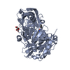
| Structure viewer | Molecule: MolmilJmol/JSmol |
|---|
- Downloads & links
-Download
| PDBx/mmCIF format | 4h3f.cif.gz | 181.6 KB | Display | PDBx/mmCIF format |
|---|---|---|---|---|
| PDB format | pdb4h3f.ent.gz | 149.6 KB | Display | PDB format |
| PDBx/mmJSON format | 4h3f.json.gz | Tree view | PDBx/mmJSON format | |
| Others |  Other downloads Other downloads |
-Validation report
| Arichive directory | https://data.pdbj.org/pub/pdb/validation_reports/h3/4h3fftp://data.pdbj.org/pub/pdb/validation_reports/h3/4h3f | HTTPS FTP |
|---|
-Related structure data


-Links
 PDBj
PDBj


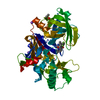
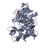
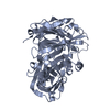
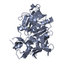
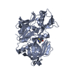
- Assembly
| Deposited unit | 
| ||||||||
|---|---|---|---|---|---|---|---|---|---|
| 1 | 
| ||||||||
| 2 | 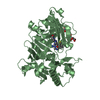
| ||||||||
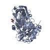
| Unit cell |
|
-Components
| #1: Protein | / Aspartyl protease 2 / ASP2 / Asp 2 / Beta-site amyloid precursor protein cleaving enzyme 1 / Beta- ...Aspartyl protease 2 / ASP2 / Asp 2 / Beta-site amyloid precursor protein cleaving enzyme 1 / Beta-site APP cleaving enzyme 1 / Memapsin-2 / Membrane-associated aspartic protease 2 Mass: 46249.926 Da / Num. of mol.: 2 / Fragment: UNP Residues 41-454 Source method: isolated from a genetically manipulated source Source: (gene. exp.) Homo sapiens (human) / Gene: BACE, BACE1, KIAA1149 / Production host:  Escherichia coli (E. coli) / Strain (production host): BL21(DE3) / References: UniProt: P56817, memapsin 2 Escherichia coli (E. coli) / Strain (production host): BL21(DE3) / References: UniProt: P56817, memapsin 2#2: Chemical | Tartaric acid  Mass: 150.087 Da / Num. of mol.: 3 / Source method: obtained synthetically / Formula: C4H6O6 Mass: 150.087 Da / Num. of mol.: 3 / Source method: obtained synthetically / Formula: C4H6O6#3: Chemical |   Mass: 458.535 Da / Num. of mol.: 2 / Source method: obtained synthetically / Formula: C24H22N6O2S Mass: 458.535 Da / Num. of mol.: 2 / Source method: obtained synthetically / Formula: C24H22N6O2S#4: Water | ChemComp-HOH / | Water Mass: 18.015 Da / Num. of mol.: 887 / Source method: isolated from a natural source / Formula: H2O Mass: 18.015 Da / Num. of mol.: 887 / Source method: isolated from a natural source / Formula: H2O |
|---|
-Experimental details
-Experiment
| Experiment | Method: X-RAY DIFFRACTION / Number of used crystals: 1 |
|---|
- Sample preparation
| Crystal | Density Matthews: 2.73 Å3/Da / Density % sol: 54.97 % |
|---|---|
| Crystal grow | Temperature: 277 K / Method: vapor diffusion, hanging drop / Details: VAPOR DIFFUSION, HANGING DROP, temperature 277K |
-Data collection
| Diffraction | Mean temperature: 95 K | |||||||||||||||||||||||||||||||||||||||||||||||||||||||||||||||||||||||||||||||||||||||||||||||||||||||||||||||||||||||||||||||||||||||||||||||||||
|---|---|---|---|---|---|---|---|---|---|---|---|---|---|---|---|---|---|---|---|---|---|---|---|---|---|---|---|---|---|---|---|---|---|---|---|---|---|---|---|---|---|---|---|---|---|---|---|---|---|---|---|---|---|---|---|---|---|---|---|---|---|---|---|---|---|---|---|---|---|---|---|---|---|---|---|---|---|---|---|---|---|---|---|---|---|---|---|---|---|---|---|---|---|---|---|---|---|---|---|---|---|---|---|---|---|---|---|---|---|---|---|---|---|---|---|---|---|---|---|---|---|---|---|---|---|---|---|---|---|---|---|---|---|---|---|---|---|---|---|---|---|---|---|---|---|---|---|---|
| Diffraction source | Source: ROTATING ANODE / Type: RIGAKU RU200 / Wavelength: 1.54178 Å | |||||||||||||||||||||||||||||||||||||||||||||||||||||||||||||||||||||||||||||||||||||||||||||||||||||||||||||||||||||||||||||||||||||||||||||||||||
| Detector | Type: RIGAKU RAXIS IV / Detector: IMAGE PLATE / Date: Apr 17, 2006 | |||||||||||||||||||||||||||||||||||||||||||||||||||||||||||||||||||||||||||||||||||||||||||||||||||||||||||||||||||||||||||||||||||||||||||||||||||
| Radiation | Protocol: SINGLE WAVELENGTH / Monochromatic (M) / Laue (L): M / Scattering type: x-ray | |||||||||||||||||||||||||||||||||||||||||||||||||||||||||||||||||||||||||||||||||||||||||||||||||||||||||||||||||||||||||||||||||||||||||||||||||||
| Radiation wavelength | Wavelength: 1.54178 Å / Relative weight: 1 | |||||||||||||||||||||||||||||||||||||||||||||||||||||||||||||||||||||||||||||||||||||||||||||||||||||||||||||||||||||||||||||||||||||||||||||||||||
| Reflection | Resolution: 1.7→20 Å / Num. all: 111855 / Num. obs: 106993 / % possible obs: 95.7 % / Observed criterion σ(F): 0 / Observed criterion σ(I): 0 / Redundancy: 2.4 % / Rmerge(I) obs: 0.065 / Χ2: 1 / Net I/σ(I): 10.7 | |||||||||||||||||||||||||||||||||||||||||||||||||||||||||||||||||||||||||||||||||||||||||||||||||||||||||||||||||||||||||||||||||||||||||||||||||||
| Reflection shell |
|
- Processing
| Software |
| ||||||||||||||||||||||||||||
|---|---|---|---|---|---|---|---|---|---|---|---|---|---|---|---|---|---|---|---|---|---|---|---|---|---|---|---|---|---|
| Refinement | Method to determine structure: FOURIER SYNTHESIS / Resolution: 1.7→20 Å / Occupancy max: 1 / Occupancy min: 1 / σ(F): 0
| ||||||||||||||||||||||||||||
| Displacement parameters | Biso max: 73.86 Å2 / Biso mean: 21.1717 Å2 / Biso min: 6.61 Å2
| ||||||||||||||||||||||||||||
| Refinement step | Cycle: LAST / Resolution: 1.7→20 Å
| ||||||||||||||||||||||||||||
| Refine LS restraints |
| ||||||||||||||||||||||||||||
| Xplor file |
|
