 Movie
Movie Controller

[English] 日本語
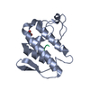
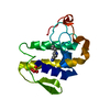
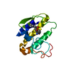
 Yorodumi
- PDB-4fga: Design of peptide inhibitors of group II phospholipase A2: Crysta... -
+ Open data

- Basic information
| Entry | Database: PDB / ID: 4fga | ||||||
|---|---|---|---|---|---|---|---|
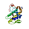
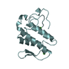
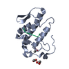
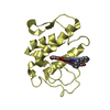
| Title | Design of peptide inhibitors of group II phospholipase A2: Crystal structure of the complex of phospholipsae A2 with a designed tripeptide, Ala- Tyr- Lys at 2.3 A resolution | ||||||
 Components Components |
| ||||||
 Keywords Keywords | HYDROLASE/HYDROLASE INHIBITOR /  PLA2 / Anti-inflammatory / Anti-coagulant / Peptide-inhibitor / Hydrolase / HYDROLASE-HYDROLASE INHIBITOR complex PLA2 / Anti-inflammatory / Anti-coagulant / Peptide-inhibitor / Hydrolase / HYDROLASE-HYDROLASE INHIBITOR complex | ||||||
| Function / homology |  Function and homology information Function and homology informationcalcium-dependent phospholipase A2 activity / phospholipase A2 / arachidonic acid secretion / phospholipid metabolic process / lipid catabolic process / negative regulation of T cell proliferation / phospholipid binding / toxin activity / calcium ion binding / extracellular regionSimilarity search - Function | ||||||
| Biological species |  Daboia russellii pulchella (snake) Daboia russellii pulchella (snake) | ||||||
| Method | X-RAY DIFFRACTION / MOLECULAR REPLACEMENT / Resolution: 2.3 Å | ||||||
 Authors Authors | Shukla, P.K. / Sinha, M. / Dey, S. / Kaur, P. / Sharma, S. / Singh, T.P. | ||||||
 Citation Citation | Journal: To be Published Title: Design of peptide inhibitors of group II phospholipase A2: Crystal structure of the complex of phospholipsae A2 with a designed tripeptide, Ala- Tyr- Lys at 2.3 A resolution Authors: Shukla, P.K. / Sinha, M. / Dey, S. / Kaur, P. / Sharma, S. / Singh, T.P. | ||||||
| History |
|
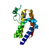
- Structure visualization
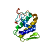
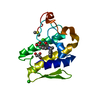
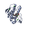
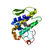
| Structure viewer | Molecule: MolmilJmol/JSmol |
|---|
- Downloads & links
-Download
| PDBx/mmCIF format | 4fga.cif.gz | 39.5 KB | Display | PDBx/mmCIF format |
|---|---|---|---|---|
| PDB format | pdb4fga.ent.gz | 26.6 KB | Display | PDB format |
| PDBx/mmJSON format | 4fga.json.gz | Tree view | PDBx/mmJSON format | |
| Others |  Other downloads Other downloads |
-Validation report
| Arichive directory | https://data.pdbj.org/pub/pdb/validation_reports/fg/4fgaftp://data.pdbj.org/pub/pdb/validation_reports/fg/4fga | HTTPS FTP |
|---|
-Related structure data
| Related structure data | 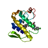 4eixS S: Starting model for refinement |
|---|---|
| Similar structure data |


-Links
 PDBj
PDBj

- Assembly
| Deposited unit | 
| ||||||||
|---|---|---|---|---|---|---|---|---|---|
| 1 |
| ||||||||
| Unit cell |
|
-Components
| #1: Protein | Mass: 13629.767 Da / Num. of mol.: 1 / Source method: isolated from a natural source / Source: (natural) Daboia russellii pulchella (snake)References: UniProt: D0VX11, UniProt: P59071*PLUS, phospholipase A2 |
|---|---|
| #2: Protein/peptide | Mass: 381.447 Da / Num. of mol.: 1 / Source method: obtained synthetically / Details: synthetic peptide |
| #3: Water | ChemComp-HOH / Water Mass: 18.015 Da / Num. of mol.: 98 / Source method: isolated from a natural source / Formula: H2O Mass: 18.015 Da / Num. of mol.: 98 / Source method: isolated from a natural source / Formula: H2O |
-Experimental details
-Experiment
| Experiment | Method: X-RAY DIFFRACTION / Number of used crystals: 1 |
|---|
- Sample preparation
| Crystal | Density Matthews: 2.45 Å3/Da / Density % sol: 49.77 % |
|---|---|
| Crystal grow | Temperature: 298 K / Method: vapor diffusion, hanging drop / pH: 7.2 Details: 0.3M AMMONIUM SULPHATE, 30% PEG 4000, pH 7.2, VAPOR DIFFUSION, HANGING DROP, temperature 298K |
-Data collection
| Diffraction | Mean temperature: 288 K |
|---|---|
| Diffraction source | Source: ROTATING ANODE / Type: RIGAKU RU300 / Wavelength: 1.54 Å |
| Detector | Type: MARRESEARCH / Detector: IMAGE PLATE / Date: Mar 7, 2006 / Details: mirror |
| Radiation | Monochromator: Graphite / Protocol: SINGLE WAVELENGTH / Monochromatic (M) / Laue (L): M / Scattering type: x-ray |
| Radiation wavelength | Wavelength: 1.54 Å / Relative weight: 1 |
| Reflection | Resolution: 2.3→53.17 Å / Num. obs: 5887 / % possible obs: 99.3 % / Observed criterion σ(F): 0 / Observed criterion σ(I): 0 / Rsym value: 0.116 / Net I/σ(I): 8.6 |
| Reflection shell | Resolution: 2.3→2.38 Å / Mean I/σ(I) obs: 2.4 / Rsym value: 0.368 / % possible all: 100 |
- Processing
| Software |
| |||||||||||||||||||||||||||||||||||||||||||||
|---|---|---|---|---|---|---|---|---|---|---|---|---|---|---|---|---|---|---|---|---|---|---|---|---|---|---|---|---|---|---|---|---|---|---|---|---|---|---|---|---|---|---|---|---|---|---|
| Refinement | Method to determine structure: MOLECULAR REPLACEMENT Starting model: 4EIX Resolution: 2.3→53.17 Å / Cor.coef. Fo:Fc: 0.913 / Cor.coef. Fo:Fc free: 0.898 / SU B: 5.781 / SU ML: 0.138 / Cross valid method: THROUGHOUT / σ(F): 0 / σ(I): 0 / ESU R: 0.413 / ESU R Free: 0.249 / Stereochemistry target values: MAXIMUM LIKELIHOOD / Details: HYDROGENS HAVE BEEN USED IF PRESENT IN THE INPUT
| |||||||||||||||||||||||||||||||||||||||||||||
| Solvent computation | Ion probe radii: 0.8 Å / Shrinkage radii: 0.8 Å / VDW probe radii: 1.2 Å / Solvent model: MASK | |||||||||||||||||||||||||||||||||||||||||||||
| Displacement parameters | Biso mean: 27.953 Å2
| |||||||||||||||||||||||||||||||||||||||||||||
| Refinement step | Cycle: LAST / Resolution: 2.3→53.17 Å
| |||||||||||||||||||||||||||||||||||||||||||||
| Refine LS restraints |
| |||||||||||||||||||||||||||||||||||||||||||||
| LS refinement shell | Resolution: 2.301→2.36 Å / Total num. of bins used: 20
|
