 Movie
Movie Controller

[English] 日本語
 Yorodumi
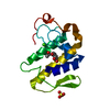
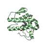
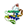
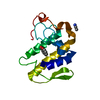
- PDB-1sv9: Crystal structure of the complex formed between groupII phospholi... -
+ Open data

- Basic information
| Entry | Database: PDB / ID: 1sv9 | ||||||
|---|---|---|---|---|---|---|---|
| Title | Crystal structure of the complex formed between groupII phospholipase A2 and anti-inflammatory agent 2-[(2,6-Dichlorophenyl)amino] benzeneacetic acid at 2.7A resolution | ||||||
 Components Components | Phospholipase A2 | ||||||
 Keywords Keywords | TOXIN / Phospholipase A2 / complex / diclofenac / 2.7A resolution | ||||||
| Function / homology |  Function and homology information Function and homology informationcalcium-dependent phospholipase A2 activity / phospholipase A2 / arachidonic acid secretion / phospholipid metabolic process / lipid catabolic process / negative regulation of T cell proliferation / phospholipid binding / toxin activity / calcium ion binding / extracellular regionSimilarity search - Function | ||||||
| Biological species |  Daboia russellii russellii (snake) Daboia russellii russellii (snake) | ||||||
| Method | X-RAY DIFFRACTION / MOLECULAR REPLACEMENT / Resolution: 2.71 Å | ||||||
 Authors Authors | Senthil kumar, R. / Singh, N. / Ethayathulla, A.S. / Prem kumar, R. / Sharma, S. / Singh, T.P. | ||||||
 Citation Citation | Journal: To be Published Title: Crystal structure of the complex formed between group II phospholipase A2 and anti-inflammatory agent 2-[(2,6-Dichlorophenyl)amino] benzeneacetic acid at 2.7A resolution Authors: Senthil kumar, R. / Singh, N. / Ethayathulla, A.S. / Prem kumar, R. / Sharma, S. / Singh, T.P. | ||||||
| History |
|
- Structure visualization
| Structure viewer | Molecule: MolmilJmol/JSmol |
|---|
- Downloads & links
-Download
| PDBx/mmCIF format | 1sv9.cif.gz | 38.4 KB | Display | PDBx/mmCIF format |
|---|---|---|---|---|
| PDB format | pdb1sv9.ent.gz | 25.7 KB | Display | PDB format |
| PDBx/mmJSON format | 1sv9.json.gz | Tree view | PDBx/mmJSON format | |
| Others |  Other downloads Other downloads |
-Validation report
| Arichive directory | https://data.pdbj.org/pub/pdb/validation_reports/sv/1sv9ftp://data.pdbj.org/pub/pdb/validation_reports/sv/1sv9 | HTTPS FTP |
|---|
-Related structure data
| Related structure data |  1fb2S S: Starting model for refinement |
|---|---|
| Similar structure data |


-Links
 PDBj
PDBj

- Assembly
| Deposited unit | 
| ||||||||
|---|---|---|---|---|---|---|---|---|---|
| 1 |
| ||||||||
| Unit cell |
| ||||||||
| Details | The biological Unit is Monomer |
-Components
| #1: Protein | / Phosphatidylcholine 2-acylhydrolase Mass: 13629.767 Da / Num. of mol.: 1 / Source method: isolated from a natural source / Source: (natural) Daboia russellii russellii (snake) / Secretion: venom / Species: Daboia russellii / Strain: russellii / References: UniProt: P59071, phospholipase A2 |
|---|---|
| #2: Chemical | ChemComp-DIF / Diclofenac  Mass: 296.149 Da / Num. of mol.: 1 / Source method: obtained synthetically / Formula: C14H11Cl2NO2 / Comment: antiinflammatory, medication*YM Mass: 296.149 Da / Num. of mol.: 1 / Source method: obtained synthetically / Formula: C14H11Cl2NO2 / Comment: antiinflammatory, medication*YM |
| #3: Water | ChemComp-HOH / Water Mass: 18.015 Da / Num. of mol.: 53 / Source method: isolated from a natural source / Formula: H2O Mass: 18.015 Da / Num. of mol.: 53 / Source method: isolated from a natural source / Formula: H2O |
-Experimental details
-Experiment
| Experiment | Method: X-RAY DIFFRACTION / Number of used crystals: 1 |
|---|
- Sample preparation
| Crystal | Density Matthews: 2.5 Å3/Da / Density % sol: 50 % |
|---|---|
| Crystal grow | Temperature: 297 K / Method: vapor diffusion, hanging drop / pH: 7.8 Details: 0.2M Ammonium sulphate, 50% PEG, pH 7.8, VAPOR DIFFUSION, HANGING DROP, temperature 297K |
-Data collection
| Diffraction | Mean temperature: 298 K |
|---|---|
| Diffraction source | Source: ROTATING ANODE / Type: RIGAKU RU300 / Wavelength: 1.5418 Å |
| Detector | Type: MARRESEARCH / Detector: IMAGE PLATE / Date: Mar 20, 2004 / Details: MIRROR |
| Radiation | Monochromator: GRAPHITE / Protocol: SINGLE WAVELENGTH / Monochromatic (M) / Laue (L): M / Scattering type: x-ray |
| Radiation wavelength | Wavelength: 1.5418 Å / Relative weight: 1 |
| Reflection | Resolution: 2.7→20 Å / Num. all: 3463 / Num. obs: 3463 / % possible obs: 90 % / Observed criterion σ(F): 0 / Observed criterion σ(I): 0 / Redundancy: 12.41 % / Biso Wilson estimate: 36.4 Å2 / Rsym value: 0.119 / Net I/σ(I): 7.3 |
| Reflection shell | Resolution: 2.7→2.76 Å / Mean I/σ(I) obs: 2.1 / Rsym value: 0.206 / % possible all: 75 |
- Processing
| Software |
| ||||||||||||||||||||||||||||||||||||
|---|---|---|---|---|---|---|---|---|---|---|---|---|---|---|---|---|---|---|---|---|---|---|---|---|---|---|---|---|---|---|---|---|---|---|---|---|---|
| Refinement | Method to determine structure: MOLECULAR REPLACEMENT Starting model: 1FB2 Resolution: 2.71→18.76 Å / Rfactor Rfree error: 0.018 / Data cutoff high absF: 774739.86 / Data cutoff low absF: 0 / Isotropic thermal model: RESTRAINED / Cross valid method: THROUGHOUT / σ(F): 0 / Stereochemistry target values: MAXIMUM LIKELIHOOD / Details: HYDROGENS HAVE BEEN ADDED IN THE RIDING POSITIONS
| ||||||||||||||||||||||||||||||||||||
| Solvent computation | Solvent model: FLAT MODEL / Bsol: 39.2549 Å2 / ksol: 0.334156 e/Å3 | ||||||||||||||||||||||||||||||||||||
| Displacement parameters | Biso mean: 27 Å2
| ||||||||||||||||||||||||||||||||||||
| Refine analyze |
| ||||||||||||||||||||||||||||||||||||
| Refinement step | Cycle: LAST / Resolution: 2.71→18.76 Å
| ||||||||||||||||||||||||||||||||||||
| Refine LS restraints |
| ||||||||||||||||||||||||||||||||||||
| LS refinement shell | Resolution: 2.7→2.87 Å / Rfactor Rfree error: 0.083 / Total num. of bins used: 6
| ||||||||||||||||||||||||||||||||||||
| Xplor file |
|
