 Movie
Movie Controller

[English] 日本語
 Yorodumi
- PDB-1jq8: Design of specific inhibitors of phospholipase A2: Crystal struct... -
+ Open data

- Basic information
| Entry | Database: PDB / ID: 1jq8 | ||||||
|---|---|---|---|---|---|---|---|
| Title | Design of specific inhibitors of phospholipase A2: Crystal structure of a complex formed between phospholipase A2 from Daboia russelli pulchella and a designed pentapeptide Leu-Ala-Ile-Tyr-Ser at 2.0 resolution | ||||||
 Components Components |
| ||||||
 Keywords Keywords | HYDROLASE/HYDROLASE INHIBITOR /  neurotoxic / designed peptide / HYDROLASE / HYDROLASE-HYDROLASE INHIBITOR COMPLEX neurotoxic / designed peptide / HYDROLASE / HYDROLASE-HYDROLASE INHIBITOR COMPLEX | ||||||
| Function / homology |  Function and homology information Function and homology informationcalcium-dependent phospholipase A2 activity / phospholipase A2 / arachidonic acid secretion / phospholipid metabolic process / lipid catabolic process / negative regulation of T cell proliferation / phospholipid binding / toxin activity / calcium ion binding / extracellular regionSimilarity search - Function | ||||||
| Biological species |  Daboia russellii pulchella (snake) Daboia russellii pulchella (snake) | ||||||
| Method | X-RAY DIFFRACTION / SYNCHROTRON / MOLECULAR REPLACEMENT / Resolution: 2 Å | ||||||
 Authors Authors | Chandra, V. / Jasti, J. / Kaur, P. / Dey, S. / Betzel, C. / Singh, T.P. | ||||||
 Citation Citation | Journal: ACTA CRYSTALLOGR.,SECT.D / Year: 2002 Title: Design of specific peptide inhibitors of phospholipase A2: structure of a complex formed between Russell's viper phospholipase A2 and a designed peptide Leu-Ala-Ile-Tyr-Ser (LAIYS). Authors: Chandra, V. / Jasti, J. / Kaur, P. / Dey, S. / Srinivasan, A. / Betzel, C.h. / Singh, T.P. #1: Journal: J.Mol.Biol. / Year: 2000Title: Three-dimensional structure of a presynaptic neurotoxic phospholipase A2 from Daboia russelli pulchella at 2.4 resolution Authors: Chandra, V. / Kaur, P. / Srinivasan, A. / Singh, T.P. #2: Journal: Acta Crystallogr.,Sect.D / Year: 2001Title: Regulation of catalytic function by molecular association: structure of phospholipase A2 from Daboia russelli pulchella (DPLA2) at 1.9 A resolution Authors: Chandra, V. / Kaur, P. / Jasti, J. / Betzel, C. / Singh, T.P. #3: Journal: J.Mol.Biol. / Year: 2002Title: First structural evidence of a specific inhibition of phospholipase A2 by alpha-tocopherol (vitamin E) and its implications in inflammation: crystal structure of the complex formed between ...Title: First structural evidence of a specific inhibition of phospholipase A2 by alpha-tocopherol (vitamin E) and its implications in inflammation: crystal structure of the complex formed between phospholipase A2 and alpha-tocopherol at 1.8 A resolution Authors: Chandra, V. / Jasti, J. / Kaur, P. / Betzel, C. / Srinivasan, A. / Singh, T.P. #4: Journal: Biochemistry / Year: 2002Title: Structural basis of phospholipase A2 inhibition for the synthesis of prostaglandins by the plant alkaloid aristolochic acid from a 1.7 A crystal structure Authors: Chandra, V. / Jasti, J. / Kaur, P. / Srinivasan, A. / Betzel, C. / Singh, T.P. | ||||||
| History |
|
- Structure visualization
| Structure viewer | Molecule: MolmilJmol/JSmol |
|---|
- Downloads & links
-Download
| PDBx/mmCIF format | 1jq8.cif.gz | 68.9 KB | Display | PDBx/mmCIF format |
|---|---|---|---|---|
| PDB format | pdb1jq8.ent.gz | 50.8 KB | Display | PDB format |
| PDBx/mmJSON format | 1jq8.json.gz | Tree view | PDBx/mmJSON format | |
| Others |  Other downloads Other downloads |
-Validation report
| Arichive directory | https://data.pdbj.org/pub/pdb/validation_reports/jq/1jq8ftp://data.pdbj.org/pub/pdb/validation_reports/jq/1jq8 | HTTPS FTP |
|---|
-Related structure data
| Related structure data |  1fe7 S: Starting model for refinement |
|---|---|
| Similar structure data |


-Links
 PDBj
PDBj


- Assembly
| Deposited unit | 
| |||||||||||||||
|---|---|---|---|---|---|---|---|---|---|---|---|---|---|---|---|---|
| 1 | 
| |||||||||||||||
| 2 | 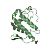
| |||||||||||||||
| Unit cell |
| |||||||||||||||
| Components on special symmetry positions |
|
-Components
| #1: Protein | Mass: 13629.767 Da / Num. of mol.: 2 / Source method: isolated from a natural source / Source: (natural) Daboia russellii pulchella (snake) / References: UniProt: P59071*PLUS, phospholipase A2#2: Protein/peptide | | Mass: 565.660 Da / Num. of mol.: 1 / Source method: obtained synthetically / Details: the sequence was Chemically synthesized. #3: Chemical | ChemComp-SO4 / | Sulfate  Mass: 96.063 Da / Num. of mol.: 1 / Source method: obtained synthetically / Formula: SO4 Mass: 96.063 Da / Num. of mol.: 1 / Source method: obtained synthetically / Formula: SO4#4: Chemical | Acetic acid  Mass: 60.052 Da / Num. of mol.: 3 / Source method: obtained synthetically / Formula: C2H4O2 Mass: 60.052 Da / Num. of mol.: 3 / Source method: obtained synthetically / Formula: C2H4O2#5: Water | ChemComp-HOH / | Water Mass: 18.015 Da / Num. of mol.: 283 / Source method: isolated from a natural source / Formula: H2O Mass: 18.015 Da / Num. of mol.: 283 / Source method: isolated from a natural source / Formula: H2OSequence details | THE COMPLETE SEQUENCE FOR THIS STRUCTURE IS 121 RESIDUES LONG. GENBANK AAB47213 (ACCESSION 1839639) ...THE COMPLETE SEQUENCE FOR THIS STRUCTURE IS 121 RESIDUES LONG. GENBANK AAB47213 (ACCESSION 1839639) MATCHED EXACTLY WITH THE FIRST 50 RESIDUES OF THIS STRUCTURE. AN ADEQUATE DATABASE MATCH COULD NOT BE FOUND FOR THE COMPLETE 121 RESIDUE SEQUENCE. THE COMPLETE SEQUENCE IS PRESENT IN THE REFERENCE PAPER: GOWDA ET AL., TOXICON 32 (6) 665-673 (1994). | |
|---|
-Experimental details
-Experiment
| Experiment | Method: X-RAY DIFFRACTION / Number of used crystals: 1 |
|---|
- Sample preparation
| Crystal | Density Matthews: 2.59 Å3/Da / Density % sol: 52.5 % | |||||||||||||||||||||||||||||||||||||||||||||||||
|---|---|---|---|---|---|---|---|---|---|---|---|---|---|---|---|---|---|---|---|---|---|---|---|---|---|---|---|---|---|---|---|---|---|---|---|---|---|---|---|---|---|---|---|---|---|---|---|---|---|---|
| Crystal grow | Temperature: 298 K / Method: vapor diffusion, hanging drop / pH: 6.5 Details: 20mM Sodium cacodylate, 1.4M Ammonium sulfate, 4mM Calcium chloride, 3% dioxane, pH 6.5, VAPOR DIFFUSION, HANGING DROP, temperature 298K | |||||||||||||||||||||||||||||||||||||||||||||||||
| Crystal grow | *PLUS Method: vapor diffusion, sitting drop | |||||||||||||||||||||||||||||||||||||||||||||||||
| Components of the solutions | *PLUS
|
-Data collection
| Diffraction | Mean temperature: 180 K |
|---|---|
| Diffraction source | Source: SYNCHROTRON / Site: EMBL/DESY, HAMBURG  / Beamline: X11 / Wavelength: 0.98 Å / Beamline: X11 / Wavelength: 0.98 Å |
| Detector | Type: MARRESEARCH / Detector: IMAGE PLATE / Date: Jun 20, 1999 |
| Radiation | Protocol: SINGLE WAVELENGTH / Monochromatic (M) / Laue (L): M / Scattering type: x-ray |
| Radiation wavelength | Wavelength: 0.98 Å / Relative weight: 1 |
| Reflection | Resolution: 2→20 Å / Num. all: 17914 / Num. obs: 17914 / % possible obs: 96.3 % / Observed criterion σ(F): 0 / Observed criterion σ(I): 0 / Redundancy: 2.7 % / Biso Wilson estimate: 31.1 Å2 / Rmerge(I) obs: 0.076 / Rsym value: 0.02 / Net I/σ(I): 9.22 |
| Reflection shell | Resolution: 2→2.07 Å / Redundancy: 2.1 % / Rmerge(I) obs: 4.3 / Mean I/σ(I) obs: 9.4 / Num. unique all: 1605 / Rsym value: 0.17 / % possible all: 90.3 |
| Reflection | *PLUS Highest resolution: 2 Å / Lowest resolution: 20 Å / Num. measured all: 222133 / Rmerge(I) obs: 0.02 |
| Reflection shell | *PLUS Highest resolution: 2 Å / % possible obs: 88.1 % / Rmerge(I) obs: 0.097 / Mean I/σ(I) obs: 2.5 |
- Processing
| Software |
| ||||||||||||||||||||||||||||||||||||||||||||||||||||||||||||||||||||||||||||||||
|---|---|---|---|---|---|---|---|---|---|---|---|---|---|---|---|---|---|---|---|---|---|---|---|---|---|---|---|---|---|---|---|---|---|---|---|---|---|---|---|---|---|---|---|---|---|---|---|---|---|---|---|---|---|---|---|---|---|---|---|---|---|---|---|---|---|---|---|---|---|---|---|---|---|---|---|---|---|---|---|---|---|
| Refinement | Method to determine structure: MOLECULAR REPLACEMENT Starting model: PDB: 1FE7 1fe7 Resolution: 2→11.94 Å / Rfactor Rfree error: 0.007 / Data cutoff high absF: 1312150.02 / Data cutoff high rms absF: 0 / Data cutoff low absF: 0 / Isotropic thermal model: RESTRAINED / Cross valid method: THROUGHOUT / σ(F): 0 / σ(I): 0 / Stereochemistry target values: Engh & Huber / Details: Used weighted full matrix least squares procedure.
| ||||||||||||||||||||||||||||||||||||||||||||||||||||||||||||||||||||||||||||||||
| Solvent computation | Solvent model: FLAT MODEL / Bsol: 77.4746 Å2 / ksol: 0.306488 e/Å3 | ||||||||||||||||||||||||||||||||||||||||||||||||||||||||||||||||||||||||||||||||
| Displacement parameters | Biso mean: 38.7 Å2
| ||||||||||||||||||||||||||||||||||||||||||||||||||||||||||||||||||||||||||||||||
| Refine analyze |
| ||||||||||||||||||||||||||||||||||||||||||||||||||||||||||||||||||||||||||||||||
| Refinement step | Cycle: LAST / Resolution: 2→11.94 Å
| ||||||||||||||||||||||||||||||||||||||||||||||||||||||||||||||||||||||||||||||||
| Refine LS restraints |
| ||||||||||||||||||||||||||||||||||||||||||||||||||||||||||||||||||||||||||||||||
| LS refinement shell | Resolution: 2→2.07 Å / Rfactor Rfree error: 0.017 / Total num. of bins used: 6
| ||||||||||||||||||||||||||||||||||||||||||||||||||||||||||||||||||||||||||||||||
| Xplor file |
| ||||||||||||||||||||||||||||||||||||||||||||||||||||||||||||||||||||||||||||||||
| Refinement | *PLUS Highest resolution: 2 Å / Lowest resolution: 20 Å / Num. reflection obs: 17914 / % reflection Rfree: 5.1 % / Rfactor Rwork: 0.189 | ||||||||||||||||||||||||||||||||||||||||||||||||||||||||||||||||||||||||||||||||
| Solvent computation | *PLUS | ||||||||||||||||||||||||||||||||||||||||||||||||||||||||||||||||||||||||||||||||
| Displacement parameters | *PLUS | ||||||||||||||||||||||||||||||||||||||||||||||||||||||||||||||||||||||||||||||||
| Refine LS restraints | *PLUS
|
