 Movie
Movie Controller

[English] 日本語
 Yorodumi
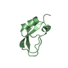
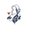
- PDB-4cvd: Crystal structure of the central repeat of cell wall binding modu... -
+ Open data

- Basic information
| Entry | Database: PDB / ID: 4cvd | ||||||
|---|---|---|---|---|---|---|---|
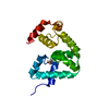
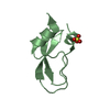
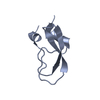
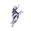
| Title | Crystal structure of the central repeat of cell wall binding module of Cpl7 | ||||||
 Components Components | LYSOZYME | ||||||
 Keywords Keywords | HYDROLASE / STREPTOCOCCUS PNEUMONIAE | ||||||
| Function / homology |  Function and homology information Function and homology informationpeptidoglycan catabolic process / cell wall macromolecule catabolic process / lysozyme / lysozyme activity / killing of cells of another organism / defense response to bacteriumSimilarity search - Function | ||||||
| Biological species |  STREPTOCOCCUS PHAGE CP-7 (virus) STREPTOCOCCUS PHAGE CP-7 (virus) | ||||||
| Method | X-RAY DIFFRACTION / SYNCHROTRON / AB INITIO PHASING / Resolution: 1.666 Å | ||||||
 Authors Authors | Silva-Martin, N. / Uson, I. / Rodriguez, D.D. / Hermoso, J.A. | ||||||
 Citation Citation | Journal: Sci Rep / Year: 2017 Title: Deciphering how Cpl-7 cell wall-binding repeats recognize the bacterial peptidoglycan. Authors: Bustamante, N. / Iglesias-Bexiga, M. / Bernardo-Garcia, N. / Silva-Martin, N. / Garcia, G. / Campanero-Rhodes, M.A. / Garcia, E. / Uson, I. / Buey, R.M. / Garcia, P. / Hermoso, J.A. / Bruix, M. / Menendez, M. | ||||||
| History |
|
- Structure visualization
| Structure viewer | Molecule: MolmilJmol/JSmol |
|---|
- Downloads & links
-Download
| PDBx/mmCIF format | 4cvd.cif.gz | 19.9 KB | Display | PDBx/mmCIF format |
|---|---|---|---|---|
| PDB format | pdb4cvd.ent.gz | 12.1 KB | Display | PDB format |
| PDBx/mmJSON format | 4cvd.json.gz | Tree view | PDBx/mmJSON format | |
| Others |  Other downloads Other downloads |
-Validation report
| Arichive directory | https://data.pdbj.org/pub/pdb/validation_reports/cv/4cvdftp://data.pdbj.org/pub/pdb/validation_reports/cv/4cvd | HTTPS FTP |
|---|
-Related structure data

-Links
 PDBj
PDBj- Assembly
| Deposited unit | 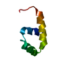
| ||||||||
|---|---|---|---|---|---|---|---|---|---|
| 1 |
| ||||||||
| Unit cell |
|
-Components
| #1: Protein/peptide | / CP-7 LYSIN / ENDOLYSIN / MURAMIDASE / CPL-7 Mass: 5263.677 Da / Num. of mol.: 1 Fragment: CENTRAL REPEAT OF CELL WALL BINDING MODULE, RESIDUES 205-252 Source method: isolated from a genetically manipulated source Source: (gene. exp.) STREPTOCOCCUS PHAGE CP-7 (virus) / Production host:  ESCHERICHIA COLI (E. coli) / References: UniProt: P19385, lysozyme ESCHERICHIA COLI (E. coli) / References: UniProt: P19385, lysozyme |
|---|---|
| #2: Water | ChemComp-HOH / Water Mass: 18.015 Da / Num. of mol.: 32 / Source method: isolated from a natural source / Formula: H2O Mass: 18.015 Da / Num. of mol.: 32 / Source method: isolated from a natural source / Formula: H2O |
-Experimental details
-Experiment
| Experiment | Method: X-RAY DIFFRACTION / Number of used crystals: 1 |
|---|
- Sample preparation
| Crystal | Density Matthews: 2.34 Å3/Da / Density % sol: 47.4 % / Description: NONE |
|---|---|
| Crystal grow | Method: vapor diffusion, hanging drop / pH: 5.5 Details: 1.3 M TRI-SODIUM CITRATE, HEGA-8 AS ADITIVE, VAPOR DIFFUSION, HANGING DROP, pH 5.5 |
-Data collection
| Diffraction | Mean temperature: 100 K |
|---|---|
| Diffraction source | Source: SYNCHROTRON / Site: ESRF  / Beamline: ID14-1 / Wavelength: 0.9334 / Beamline: ID14-1 / Wavelength: 0.9334 |
| Detector | Type: ADSC QUANTUM 210 / Detector: CCD / Date: Sep 23, 2010 / Details: MIRRORS |
| Radiation | Protocol: SINGLE WAVELENGTH / Monochromatic (M) / Laue (L): M / Scattering type: x-ray |
| Radiation wavelength | Wavelength: 0.9334 Å / Relative weight: 1 |
| Reflection | Resolution: 1.67→28.54 Å / Num. obs: 4935 / % possible obs: 99.7 % / Observed criterion σ(I): 2 / Redundancy: 10.7 % / Biso Wilson estimate: 12.17 Å2 / Rmerge(I) obs: 0.05 / Net I/σ(I): 10.3 |
| Reflection shell | Resolution: 1.67→1.76 Å / Redundancy: 10.4 % / Rmerge(I) obs: 0.18 / Mean I/σ(I) obs: 4.3 / % possible all: 98.7 |
- Processing
| Software |
| ||||||||||||||||||||||||
|---|---|---|---|---|---|---|---|---|---|---|---|---|---|---|---|---|---|---|---|---|---|---|---|---|---|
| Refinement | Method to determine structure: AB INITIO PHASING Starting model: NONE Resolution: 1.666→25.2 Å / SU ML: 0.18 / σ(F): 1.54 / Phase error: 19.78 / Stereochemistry target values: ML
| ||||||||||||||||||||||||
| Solvent computation | Shrinkage radii: 0.9 Å / VDW probe radii: 1.11 Å / Solvent model: FLAT BULK SOLVENT MODEL | ||||||||||||||||||||||||
| Displacement parameters | Biso mean: 11 Å2 | ||||||||||||||||||||||||
| Refinement step | Cycle: LAST / Resolution: 1.666→25.2 Å
| ||||||||||||||||||||||||
| Refine LS restraints |
| ||||||||||||||||||||||||
| LS refinement shell |
|
