 Movie
Movie Controller

[English] 日本語
 Yorodumi
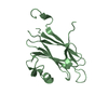
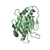
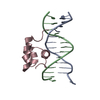
- PDB-5i8l: Crystal structure of the full-length cell wall-binding module of ... -
+ Open data

- Basic information
| Entry | Database: PDB / ID: 5i8l | ||||||
|---|---|---|---|---|---|---|---|
| Title | Crystal structure of the full-length cell wall-binding module of Cpl7 mutant R223A | ||||||
 Components Components | Lysozyme | ||||||
 Keywords Keywords | HYDROLASE / cell-wall binding domain | ||||||
| Function / homology |  Function and homology information Function and homology informationpeptidoglycan catabolic process / cell wall macromolecule catabolic process / lysozyme / lysozyme activity / killing of cells of another organism / defense response to bacteriumSimilarity search - Function | ||||||
| Biological species |  Streptococcus phage Cp-7 (virus) Streptococcus phage Cp-7 (virus) | ||||||
| Method | X-RAY DIFFRACTION / SYNCHROTRON / MOLECULAR REPLACEMENT / Resolution: 2.801 Å | ||||||
 Authors Authors | Bernardo-Garcia, N. / Silva-Martin, N. / Uson, I. / Hermoso, J.A. | ||||||
 Citation Citation | Journal: Sci Rep / Year: 2017 Title: Deciphering how Cpl-7 cell wall-binding repeats recognize the bacterial peptidoglycan. Authors: Bustamante, N. / Iglesias-Bexiga, M. / Bernardo-Garcia, N. / Silva-Martin, N. / Garcia, G. / Campanero-Rhodes, M.A. / Garcia, E. / Uson, I. / Buey, R.M. / Garcia, P. / Hermoso, J.A. / Bruix, M. / Menendez, M. | ||||||
| History |
|
- Structure visualization
| Structure viewer | Molecule: MolmilJmol/JSmol |
|---|
- Downloads & links
-Download
| PDBx/mmCIF format | 5i8l.cif.gz | 90.7 KB | Display | PDBx/mmCIF format |
|---|---|---|---|---|
| PDB format | pdb5i8l.ent.gz | 70.7 KB | Display | PDB format |
| PDBx/mmJSON format | 5i8l.json.gz | Tree view | PDBx/mmJSON format | |
| Others |  Other downloads Other downloads |
-Validation report
| Arichive directory | https://data.pdbj.org/pub/pdb/validation_reports/i8/5i8lftp://data.pdbj.org/pub/pdb/validation_reports/i8/5i8l | HTTPS FTP |
|---|
-Related structure data
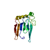
| Related structure data | 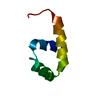 4cvdSC S: Starting model for refinement C: citing same article ( |
|---|---|
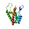
| Similar structure data |
-Links
 PDBj
PDBj- Assembly
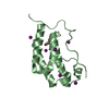
| Deposited unit | 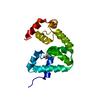
| ||||||||
|---|---|---|---|---|---|---|---|---|---|
| 1 |
| ||||||||
| Unit cell |
|
-Components
| #1: Protein | / CP-7 lysin / Endolysin / Muramidase Mass: 15617.854 Da / Num. of mol.: 1 / Mutation: yes Source method: isolated from a genetically manipulated source Source: (gene. exp.) Streptococcus phage Cp-7 (virus) / Gene: CPL7 / Production host:  Escherichia coli BL21 (bacteria) / References: UniProt: P19385, lysozyme Escherichia coli BL21 (bacteria) / References: UniProt: P19385, lysozyme |
|---|---|
| #2: Chemical | ChemComp-GOL / Glycerol  Mass: 92.094 Da / Num. of mol.: 1 / Source method: obtained synthetically / Formula: C3H8O3 Mass: 92.094 Da / Num. of mol.: 1 / Source method: obtained synthetically / Formula: C3H8O3 |
| #3: Water | ChemComp-HOH / Water Mass: 18.015 Da / Num. of mol.: 6 / Source method: isolated from a natural source / Formula: H2O Mass: 18.015 Da / Num. of mol.: 6 / Source method: isolated from a natural source / Formula: H2O |
-Experimental details
-Experiment
| Experiment | Method: X-RAY DIFFRACTION / Number of used crystals: 1 |
|---|
- Sample preparation
| Crystal | Density Matthews: 2.05 Å3/Da / Density % sol: 39.95 % |
|---|---|
| Crystal grow | Temperature: 291 K / Method: vapor diffusion, sitting drop / Details: 1.1 M tri-Sodium citrate |
-Data collection
| Diffraction | Mean temperature: 100 K |
|---|---|
| Diffraction source | Source: SYNCHROTRON / Site: ALBA  / Beamline: XALOC / Wavelength: 1.05793 Å / Beamline: XALOC / Wavelength: 1.05793 Å |
| Detector | Type: DECTRIS PILATUS 6M / Detector: PIXEL / Date: Feb 7, 2014 |
| Radiation | Protocol: SINGLE WAVELENGTH / Monochromatic (M) / Laue (L): M / Scattering type: x-ray |
| Radiation wavelength | Wavelength: 1.05793 Å / Relative weight: 1 |
| Reflection | Resolution: 2.8→43.04 Å / Num. obs: 2800 / % possible obs: 81.2 % / Redundancy: 3.9 % / Rmerge(I) obs: 0.075 / Net I/σ(I): 10.8 |
| Reflection shell | Resolution: 2.8→2.95 Å / Redundancy: 3.7 % / Rmerge(I) obs: 0.15 / Mean I/σ(I) obs: 6.3 / % possible all: 90.8 |
- Processing
| Software |
| |||||||||||||||||||||||||||||||||||||||||||||||||||||||||||||||||||||||||||||||||||||||||||||||||||||||||||||||||||||||||||||||||||||||||||||||||||||||||||||||||||||||||||||||||||||||||||||||||||||||||||||||||||||||||||||||||
|---|---|---|---|---|---|---|---|---|---|---|---|---|---|---|---|---|---|---|---|---|---|---|---|---|---|---|---|---|---|---|---|---|---|---|---|---|---|---|---|---|---|---|---|---|---|---|---|---|---|---|---|---|---|---|---|---|---|---|---|---|---|---|---|---|---|---|---|---|---|---|---|---|---|---|---|---|---|---|---|---|---|---|---|---|---|---|---|---|---|---|---|---|---|---|---|---|---|---|---|---|---|---|---|---|---|---|---|---|---|---|---|---|---|---|---|---|---|---|---|---|---|---|---|---|---|---|---|---|---|---|---|---|---|---|---|---|---|---|---|---|---|---|---|---|---|---|---|---|---|---|---|---|---|---|---|---|---|---|---|---|---|---|---|---|---|---|---|---|---|---|---|---|---|---|---|---|---|---|---|---|---|---|---|---|---|---|---|---|---|---|---|---|---|---|---|---|---|---|---|---|---|---|---|---|---|---|---|---|---|---|---|---|---|---|---|---|---|---|---|---|---|---|---|---|---|---|
| Refinement | Method to determine structure: MOLECULAR REPLACEMENT Starting model: 4cvd Resolution: 2.801→14.873 Å / SU ML: 0.26 / Cross valid method: FREE R-VALUE / σ(F): 1.35 / Phase error: 29.92
| |||||||||||||||||||||||||||||||||||||||||||||||||||||||||||||||||||||||||||||||||||||||||||||||||||||||||||||||||||||||||||||||||||||||||||||||||||||||||||||||||||||||||||||||||||||||||||||||||||||||||||||||||||||||||||||||||
| Solvent computation | Shrinkage radii: 0.9 Å / VDW probe radii: 1.11 Å | |||||||||||||||||||||||||||||||||||||||||||||||||||||||||||||||||||||||||||||||||||||||||||||||||||||||||||||||||||||||||||||||||||||||||||||||||||||||||||||||||||||||||||||||||||||||||||||||||||||||||||||||||||||||||||||||||
| Refinement step | Cycle: LAST / Resolution: 2.801→14.873 Å
| |||||||||||||||||||||||||||||||||||||||||||||||||||||||||||||||||||||||||||||||||||||||||||||||||||||||||||||||||||||||||||||||||||||||||||||||||||||||||||||||||||||||||||||||||||||||||||||||||||||||||||||||||||||||||||||||||
| Refine LS restraints |
| |||||||||||||||||||||||||||||||||||||||||||||||||||||||||||||||||||||||||||||||||||||||||||||||||||||||||||||||||||||||||||||||||||||||||||||||||||||||||||||||||||||||||||||||||||||||||||||||||||||||||||||||||||||||||||||||||
| LS refinement shell | Resolution: 2.6→2.8 Å
| |||||||||||||||||||||||||||||||||||||||||||||||||||||||||||||||||||||||||||||||||||||||||||||||||||||||||||||||||||||||||||||||||||||||||||||||||||||||||||||||||||||||||||||||||||||||||||||||||||||||||||||||||||||||||||||||||
| Refinement TLS params. | Method: refined / Refine-ID: X-RAY DIFFRACTION
| |||||||||||||||||||||||||||||||||||||||||||||||||||||||||||||||||||||||||||||||||||||||||||||||||||||||||||||||||||||||||||||||||||||||||||||||||||||||||||||||||||||||||||||||||||||||||||||||||||||||||||||||||||||||||||||||||
| Refinement TLS group |
|
