 Movie
Movie Controller

[English] 日本語
 Yorodumi
- PDB-3auc: A simplified BPTI variant with poly SER (C5S) amino acid tag at t... -
+ Open data

- Basic information
| Entry | Database: PDB / ID: 3auc | ||||||
|---|---|---|---|---|---|---|---|
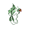
| Title | A simplified BPTI variant with poly SER (C5S) amino acid tag at the c-terminus | ||||||
 Components Components | Bovine pancreatic trypsin inhibitor Aprotinin Aprotinin | ||||||
 Keywords Keywords | HYDROLASE INHIBITOR / Serine protease inhibitor / inhibits serine protease / trypsin | ||||||
| Function / homology | Pancreatic trypsin inhibitor Kunitz domain / Factor Xa Inhibitor / Few Secondary Structures / Irregular Function and homology information Function and homology information | ||||||
| Biological species |  Bos taurus (cattle) Bos taurus (cattle) | ||||||
| Method | X-RAY DIFFRACTION / SYNCHROTRON / MOLECULAR REPLACEMENT / Resolution: 1.91 Å | ||||||
 Authors Authors | Islam, M.M. / Kato, A. / Khan, M.M.A. / Noguchi, K. / Yohda, M. / Kidokoro, S.I. / Kuroda, Y. | ||||||
 Citation Citation | Journal: To be Published Title: Effect of amino acid mutations on protein's solubility, function and structure characterized using short poly amino acid peptide tags Authors: Islam, M.M. / Kato, A. / Khan, M.M.A. / Noguchi, K. / Yohda, M. / Kidokoro, S.I. / Kuroda, Y. | ||||||
| History |
|
- Structure visualization
| Structure viewer | Molecule: MolmilJmol/JSmol |
|---|
- Downloads & links
-Download
| PDBx/mmCIF format | 3auc.cif.gz | 55.8 KB | Display | PDBx/mmCIF format |
|---|---|---|---|---|
| PDB format | pdb3auc.ent.gz | 40.9 KB | Display | PDB format |
| PDBx/mmJSON format | 3auc.json.gz | Tree view | PDBx/mmJSON format | |
| Others |  Other downloads Other downloads |
-Validation report
| Arichive directory | https://data.pdbj.org/pub/pdb/validation_reports/au/3aucftp://data.pdbj.org/pub/pdb/validation_reports/au/3auc | HTTPS FTP |
|---|
-Related structure data
| Related structure data |  3audC 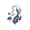 3aueC 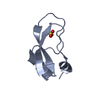 3auhC  3auiC 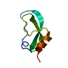 4ptiS C: citing same article ( S: Starting model for refinement |
|---|---|
| Similar structure data |


-Links
 PDBj
PDBj
- Assembly
| Deposited unit | 
| ||||||||
|---|---|---|---|---|---|---|---|---|---|
| 1 | 
| ||||||||
| 2 | 
| ||||||||
| Unit cell |
| ||||||||
| Components on special symmetry positions |
|
-Components
| #1: Protein | Aprotinin / BPTI Mass: 6566.340 Da / Num. of mol.: 2 / Mutation: A14G, A38V Source method: isolated from a genetically manipulated source Source: (gene. exp.) Bos taurus (cattle) / Cell line (production host): JM109(DE3)PLYSS / Cellular location (production host): Inclusion body / Production host:  Escherichia Coli (E. coli) Escherichia Coli (E. coli)#2: Chemical | Sulfate  Mass: 96.063 Da / Num. of mol.: 3 / Source method: obtained synthetically / Formula: SO4 Mass: 96.063 Da / Num. of mol.: 3 / Source method: obtained synthetically / Formula: SO4#3: Water | ChemComp-HOH / | Water Mass: 18.015 Da / Num. of mol.: 98 / Source method: isolated from a natural source / Formula: H2O Mass: 18.015 Da / Num. of mol.: 98 / Source method: isolated from a natural source / Formula: H2OSequence details | THIS SEQUENCE IS A SIMPLIFIED BPTI VARIANT WITH 21 ALANINES, AND IT HAS BEEN STABILIZED WITH THE ...THIS SEQUENCE IS A SIMPLIFIED | |
|---|
-Experimental details
-Experiment
| Experiment | Method: X-RAY DIFFRACTION / Number of used crystals: 1 |
|---|
- Sample preparation
| Crystal | Density Matthews: 2.05 Å3/Da / Density % sol: 40.07 % |
|---|---|
| Crystal grow | Temperature: 293 K / Method: vapor diffusion, hanging drop / pH: 8.5 Details: 30% PEG4000, 0.2M Lithium sulfate, 0.1M TrisHCl, pH 8.5, VAPOR DIFFUSION, HANGING DROP, temperature 293K |
-Data collection
| Diffraction | Mean temperature: 95 K |
|---|---|
| Diffraction source | Source: SYNCHROTRON / Site: Photon Factory  / Beamline: AR-NW12A / Wavelength: 1 Å / Beamline: AR-NW12A / Wavelength: 1 Å |
| Detector | Type: ADSC QUANTUM 210 / Detector: CCD / Date: Jan 30, 2009 |
| Radiation | Monochromator: SILICON / Protocol: SINGLE WAVELENGTH / Monochromatic (M) / Laue (L): M / Scattering type: x-ray |
| Radiation wavelength | Wavelength: 1 Å / Relative weight: 1 |
| Reflection | Resolution: 1.91→30.401 Å / Num. all: 8269 / Num. obs: 8269 / % possible obs: 96.2 % / Observed criterion σ(F): 3 / Observed criterion σ(I): 3 / Redundancy: 3.2 % / Rmerge(I) obs: 0.227 / Rsym value: 0.213 |
| Reflection shell | Resolution: 1.91→2 Å / Redundancy: 3.2 % / Rmerge(I) obs: 0.227 / Num. unique all: 8369 / % possible all: 96.2 |
- Processing
| Software |
| ||||||||||||||||||||||||||||||||||||||||||||||||||||||||||||||||||||||||||||||||||||||||||
|---|---|---|---|---|---|---|---|---|---|---|---|---|---|---|---|---|---|---|---|---|---|---|---|---|---|---|---|---|---|---|---|---|---|---|---|---|---|---|---|---|---|---|---|---|---|---|---|---|---|---|---|---|---|---|---|---|---|---|---|---|---|---|---|---|---|---|---|---|---|---|---|---|---|---|---|---|---|---|---|---|---|---|---|---|---|---|---|---|---|---|---|
| Refinement | Method to determine structure: MOLECULAR REPLACEMENT Starting model: 4PTI Resolution: 1.91→30.4 Å / Cor.coef. Fo:Fc: 0.959 / Cor.coef. Fo:Fc free: 0.924 / SU B: 9.119 / SU ML: 0.138 / Cross valid method: THROUGHOUT / σ(F): 3 / ESU R Free: 0.176 / Stereochemistry target values: MAXIMUM LIKELIHOOD
| ||||||||||||||||||||||||||||||||||||||||||||||||||||||||||||||||||||||||||||||||||||||||||
| Solvent computation | Ion probe radii: 0.8 Å / Shrinkage radii: 0.8 Å / VDW probe radii: 1.2 Å / Solvent model: MASK | ||||||||||||||||||||||||||||||||||||||||||||||||||||||||||||||||||||||||||||||||||||||||||
| Displacement parameters | Biso mean: 28.033 Å2
| ||||||||||||||||||||||||||||||||||||||||||||||||||||||||||||||||||||||||||||||||||||||||||
| Refinement step | Cycle: LAST / Resolution: 1.91→30.4 Å
| ||||||||||||||||||||||||||||||||||||||||||||||||||||||||||||||||||||||||||||||||||||||||||
| Refine LS restraints |
| ||||||||||||||||||||||||||||||||||||||||||||||||||||||||||||||||||||||||||||||||||||||||||
| LS refinement shell | Resolution: 1.913→2 Å / Total num. of bins used: 20
|
