 Movie
Movie Controller

[English] 日本語
 Yorodumi
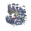
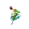
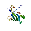
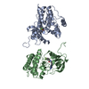
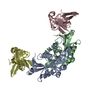
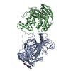
- PDB-3w1g: Crystal Structure of Human DNA ligase IV-Artemis Complex (Native) -
+ Open data

- Basic information
| Entry | Database: PDB / ID: 3w1g | ||||||
|---|---|---|---|---|---|---|---|
| Title | Crystal Structure of Human DNA ligase IV-Artemis Complex (Native) | ||||||
 Components Components |
| ||||||
 Keywords Keywords |  LIGASE / DNA ligase / non homologous end joining / DNA repair / XRCC4 LIGASE / DNA ligase / non homologous end joining / DNA repair / XRCC4 | ||||||
| Function / homology |  Function and homology information Function and homology informationDNA ligation involved in DNA recombination / positive regulation of chromosome organization / DNA ligase IV complex / DNA ligation involved in DNA repair / DNA ligase activity / DN2 thymocyte differentiation / DNA ligase (ATP) / T cell receptor V(D)J recombination / pro-B cell differentiation / DNA ligase (ATP) activity ...DNA ligation involved in DNA recombination / positive regulation of chromosome organization / DNA ligase IV complex / DNA ligation involved in DNA repair / DNA ligase activity / DN2 thymocyte differentiation / DNA ligase (ATP) / T cell receptor V(D)J recombination / pro-B cell differentiation / DNA ligase (ATP) activity / single-stranded DNA endodeoxyribonuclease activity / DNA-dependent protein kinase-DNA ligase 4 complex / single strand break repair / immunoglobulin V(D)J recombination / nonhomologous end joining complex / 5'-3' exonuclease activity / DNA ligation / V(D)J recombination / double-strand break repair via classical nonhomologous end joining / isotype switching / nucleotide-excision repair, DNA gap filling / positive regulation of neurogenesis / 5'-3' DNA exonuclease activity / response to ionizing radiation / DNA biosynthetic process / cellular response to lithium ion / 2-LTR circle formation / somatic stem cell population maintenance / ligase activity / response to X-ray / chromosome organization / interstrand cross-link repair / condensed chromosome / telomere maintenance / neurogenesis / B cell differentiation / central nervous system development / stem cell proliferation / cellular response to ionizing radiation / response to gamma radiation / Nonhomologous End-Joining (NHEJ) / double-strand break repair via nonhomologous end joining / establishment of integrated proviral latency / double-strand break repair / positive regulation of fibroblast proliferation / T cell differentiation in thymus / fibroblast proliferation / endonuclease activity / neuron apoptotic process / in utero embryonic development / adaptive immune response / negative regulation of neuron apoptotic process / cell population proliferation / damaged DNA binding / chromosome, telomeric region / Hydrolases; Acting on ester bonds / cell cycle / cell division / intracellular membrane-bounded organelle / Golgi apparatus / magnesium ion binding / DNA binding / nucleoplasm / ATP binding / nucleusSimilarity search - Function | ||||||
| Biological species |  Homo sapiens (human) Homo sapiens (human) | ||||||
| Method | X-RAY DIFFRACTION / SYNCHROTRON / MOLECULAR REPLACEMENT / Resolution: 2.55 Å | ||||||
 Authors Authors | Ochi, T. / Blundell, T.L. | ||||||
 Citation Citation | Journal: Structure / Year: 2013 Title: Structure of the catalytic region of DNA ligase IV in complex with an artemis fragment sheds light on double-strand break repair Authors: Ochi, T. / Gu, X. / Blundell, T.L. | ||||||
| History |
|
- Structure visualization
| Structure viewer | Molecule: MolmilJmol/JSmol |
|---|
- Downloads & links
-Download
| PDBx/mmCIF format | 3w1g.cif.gz | 260.3 KB | Display | PDBx/mmCIF format |
|---|---|---|---|---|
| PDB format | pdb3w1g.ent.gz | 208.1 KB | Display | PDB format |
| PDBx/mmJSON format | 3w1g.json.gz | Tree view | PDBx/mmJSON format | |
| Others |  Other downloads Other downloads |
-Validation report
| Arichive directory | https://data.pdbj.org/pub/pdb/validation_reports/w1/3w1gftp://data.pdbj.org/pub/pdb/validation_reports/w1/3w1g | HTTPS FTP |
|---|
-Related structure data
| Related structure data | 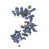 3w1bSC 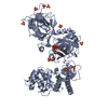 3w5oC S: Starting model for refinement C: citing same article ( |
|---|---|
| Similar structure data |


-Links
 PDBj
PDBj


- Assembly
| Deposited unit | 
| ||||||||
|---|---|---|---|---|---|---|---|---|---|
| 1 |
| ||||||||
| Unit cell |
|
-Components
| #1: Protein | / DNA ligase IV / Polydeoxyribonucleotide synthase [ATP] 4 Mass: 69538.312 Da / Num. of mol.: 1 / Fragment: Catalytic region, UNP residues 1-609 Source method: isolated from a genetically manipulated source Details: Hippocampus / Source: (gene. exp.) Homo sapiens (human) / Gene: LIG4 / Plasmid: pOPINS / Production host:  Escherichia coli (E. coli) / Strain (production host): Rosetta2(DE3)pLysS / References: UniProt: P49917, DNA ligase (ATP) Escherichia coli (E. coli) / Strain (production host): Rosetta2(DE3)pLysS / References: UniProt: P49917, DNA ligase (ATP) | ||
|---|---|---|---|
| #2: Protein/peptide | Mass: 1452.653 Da / Num. of mol.: 1 / Source method: obtained synthetically / Source: (synth.) Homo sapiens (human) / References: UniProt: Q96SD1 | ||
| #3: Chemical | ChemComp-ATP / Adenosine triphosphate  Mass: 507.181 Da / Num. of mol.: 1 / Source method: obtained synthetically / Formula: C10H16N5O13P3 / Comment: ATP, energy-carrying molecule*YM Mass: 507.181 Da / Num. of mol.: 1 / Source method: obtained synthetically / Formula: C10H16N5O13P3 / Comment: ATP, energy-carrying molecule*YM | ||
| #4: Chemical | ChemComp-SO4 / Sulfate  Mass: 96.063 Da / Num. of mol.: 15 / Source method: obtained synthetically / Formula: SO4 Mass: 96.063 Da / Num. of mol.: 15 / Source method: obtained synthetically / Formula: SO4#5: Water | ChemComp-HOH / | Water Mass: 18.015 Da / Num. of mol.: 71 / Source method: isolated from a natural source / Formula: H2O Mass: 18.015 Da / Num. of mol.: 71 / Source method: isolated from a natural source / Formula: H2O |
-Experimental details
-Experiment
| Experiment | Method: X-RAY DIFFRACTION / Number of used crystals: 1 |
|---|
- Sample preparation
| Crystal | Density Matthews: 3.07 Å3/Da / Density % sol: 59.94 % |
|---|---|
| Crystal grow | Temperature: 291 K / Method: vapor diffusion, hanging drop / pH: 5.6 Details: 2M ammonium sulfate, 10mM YCl, 100mM MES, pH 5.6, VAPOR DIFFUSION, HANGING DROP, temperature 291K |
-Data collection
| Diffraction | Mean temperature: 100 K |
|---|---|
| Diffraction source | Source: SYNCHROTRON / Site: Diamond  / Beamline: I04 / Wavelength: 0.9795 Å / Beamline: I04 / Wavelength: 0.9795 Å |
| Detector | Type: ADSC QUANTUM 315r / Detector: CCD / Date: Oct 10, 2012 |
| Radiation | Monochromator: Double crystal / Protocol: SINGLE WAVELENGTH / Monochromatic (M) / Laue (L): M / Scattering type: x-ray |
| Radiation wavelength | Wavelength: 0.9795 Å / Relative weight: 1 |
| Reflection | Resolution: 2.55→68.26 Å / Num. all: 28941 / Num. obs: 27784 / % possible obs: 96 % / Observed criterion σ(I): 2 / Redundancy: 2.8 % / Biso Wilson estimate: 46.91 Å2 / Rmerge(I) obs: 0.085 / Rsym value: 0.085 / Net I/σ(I): 8.8 |
| Reflection shell | Resolution: 2.55→2.69 Å / Redundancy: 2.9 % / Rmerge(I) obs: 0.562 / Mean I/σ(I) obs: 2 / % possible all: 97.5 |
- Processing
| Software |
| ||||||||||||||||||||||||||||||||||||||||||||||||||||||||||||||||||||||||||||||||||||||||||||||||||||||||||||||||||||||||||||||||||||||||||||||||||||||||||||||||||||||||||||||||||||||||||||||||||||||||
|---|---|---|---|---|---|---|---|---|---|---|---|---|---|---|---|---|---|---|---|---|---|---|---|---|---|---|---|---|---|---|---|---|---|---|---|---|---|---|---|---|---|---|---|---|---|---|---|---|---|---|---|---|---|---|---|---|---|---|---|---|---|---|---|---|---|---|---|---|---|---|---|---|---|---|---|---|---|---|---|---|---|---|---|---|---|---|---|---|---|---|---|---|---|---|---|---|---|---|---|---|---|---|---|---|---|---|---|---|---|---|---|---|---|---|---|---|---|---|---|---|---|---|---|---|---|---|---|---|---|---|---|---|---|---|---|---|---|---|---|---|---|---|---|---|---|---|---|---|---|---|---|---|---|---|---|---|---|---|---|---|---|---|---|---|---|---|---|---|---|---|---|---|---|---|---|---|---|---|---|---|---|---|---|---|---|---|---|---|---|---|---|---|---|---|---|---|---|---|---|---|---|
| Refinement | Method to determine structure: MOLECULAR REPLACEMENT Starting model: PDB ENTRY 3W1B Resolution: 2.55→59.462 Å / Occupancy max: 1 / Occupancy min: 0.47 / SU ML: 0.33 / σ(F): 1.35 / Phase error: 23.68 / Stereochemistry target values: ML
| ||||||||||||||||||||||||||||||||||||||||||||||||||||||||||||||||||||||||||||||||||||||||||||||||||||||||||||||||||||||||||||||||||||||||||||||||||||||||||||||||||||||||||||||||||||||||||||||||||||||||
| Solvent computation | Shrinkage radii: 0.9 Å / VDW probe radii: 1.11 Å / Solvent model: FLAT BULK SOLVENT MODEL | ||||||||||||||||||||||||||||||||||||||||||||||||||||||||||||||||||||||||||||||||||||||||||||||||||||||||||||||||||||||||||||||||||||||||||||||||||||||||||||||||||||||||||||||||||||||||||||||||||||||||
| Displacement parameters | Biso max: 178.26 Å2 / Biso mean: 59.456 Å2 / Biso min: 22.45 Å2 | ||||||||||||||||||||||||||||||||||||||||||||||||||||||||||||||||||||||||||||||||||||||||||||||||||||||||||||||||||||||||||||||||||||||||||||||||||||||||||||||||||||||||||||||||||||||||||||||||||||||||
| Refinement step | Cycle: LAST / Resolution: 2.55→59.462 Å
| ||||||||||||||||||||||||||||||||||||||||||||||||||||||||||||||||||||||||||||||||||||||||||||||||||||||||||||||||||||||||||||||||||||||||||||||||||||||||||||||||||||||||||||||||||||||||||||||||||||||||
| Refine LS restraints |
| ||||||||||||||||||||||||||||||||||||||||||||||||||||||||||||||||||||||||||||||||||||||||||||||||||||||||||||||||||||||||||||||||||||||||||||||||||||||||||||||||||||||||||||||||||||||||||||||||||||||||
| LS refinement shell | Refine-ID: X-RAY DIFFRACTION / Total num. of bins used: 10
| ||||||||||||||||||||||||||||||||||||||||||||||||||||||||||||||||||||||||||||||||||||||||||||||||||||||||||||||||||||||||||||||||||||||||||||||||||||||||||||||||||||||||||||||||||||||||||||||||||||||||
| Refinement TLS params. | Method: refined / Refine-ID: X-RAY DIFFRACTION
| ||||||||||||||||||||||||||||||||||||||||||||||||||||||||||||||||||||||||||||||||||||||||||||||||||||||||||||||||||||||||||||||||||||||||||||||||||||||||||||||||||||||||||||||||||||||||||||||||||||||||
| Refinement TLS group |
|
