 Movie
Movie Controller

[English] 日本語
 Yorodumi
- PDB-3lv4: Crystal structure of the glycoside hydrolase, family 43 YxiA prot... -
+ Open data

- Basic information
| Entry | Database: PDB / ID: 3lv4 | ||||||
|---|---|---|---|---|---|---|---|
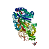
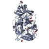
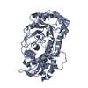
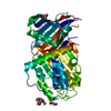
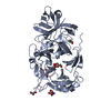
| Title | Crystal structure of the glycoside hydrolase, family 43 YxiA protein from Bacillus licheniformis. Northeast Structural Genomics Consortium Target BiR14. | ||||||
 Components Components | Glycoside hydrolase YxiA | ||||||
 Keywords Keywords |  HYDROLASE / Glycoside hydrolase / similar to arabinan endo-1 / 5-alpha-L-arabinosidase / NESG / Structural Genomics / PSI-2 / Protein Structure Initiative / Northeast Structural Genomics Consortium HYDROLASE / Glycoside hydrolase / similar to arabinan endo-1 / 5-alpha-L-arabinosidase / NESG / Structural Genomics / PSI-2 / Protein Structure Initiative / Northeast Structural Genomics Consortium | ||||||
| Function / homology |  Function and homology information Function and homology informationhydrolase activity, hydrolyzing O-glycosyl compounds / carbohydrate metabolic process / metal ion bindingSimilarity search - Function | ||||||
| Biological species |  Bacillus licheniformis (bacteria) Bacillus licheniformis (bacteria) | ||||||
| Method | X-RAY DIFFRACTION / SYNCHROTRON / SAD / Resolution: 1.695 Å | ||||||
 Authors Authors | Vorobiev, S. / Abashidze, M. / Seetharaman, J. / Belote, R.L. / Ciccosanti, C. / Sahdev, S. / Xiao, R. / Acton, T.B. / Everett, J.K. / Montelione, G.T. ...Vorobiev, S. / Abashidze, M. / Seetharaman, J. / Belote, R.L. / Ciccosanti, C. / Sahdev, S. / Xiao, R. / Acton, T.B. / Everett, J.K. / Montelione, G.T. / Tong, L. / Hunt, J.F. / Northeast Structural Genomics Consortium (NESG) | ||||||
 Citation Citation | Journal: To be Published Title: Crystal structure of the glycoside hydrolase, family 43 YxiA protein from Bacillus licheniformis. Authors: Vorobiev, S. / Abashidze, M. / Seetharaman, J. / Belote, R.L. / Ciccosanti, C. / Sahdev, S. / Xiao, R. / Acton, T.B. / Everett, J.K. / Montelione, G.T. / Tong, L. / Hunt, J.F. | ||||||
| History |
|
- Structure visualization
| Structure viewer | Molecule: MolmilJmol/JSmol |
|---|
- Downloads & links
-Download
| PDBx/mmCIF format | 3lv4.cif.gz | 360.7 KB | Display | PDBx/mmCIF format |
|---|---|---|---|---|
| PDB format | pdb3lv4.ent.gz | 305.2 KB | Display | PDB format |
| PDBx/mmJSON format | 3lv4.json.gz | Tree view | PDBx/mmJSON format | |
| Others |  Other downloads Other downloads |
-Validation report
| Arichive directory | https://data.pdbj.org/pub/pdb/validation_reports/lv/3lv4ftp://data.pdbj.org/pub/pdb/validation_reports/lv/3lv4 | HTTPS FTP |
|---|
-Related structure data
| Similar structure data | |
|---|---|
| Other databases |


-Links
 PDBj
PDBj- Assembly
| Deposited unit | 
| ||||||||
|---|---|---|---|---|---|---|---|---|---|
| 1 | 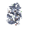
| ||||||||
| 2 | 
| ||||||||
| 3 | 
| ||||||||
| Unit cell |
| ||||||||
| Details | monomer according to gel filtration |
-Components
| #1: Protein | Mass: 51781.039 Da / Num. of mol.: 2 Source method: isolated from a genetically manipulated source Source: (gene. exp.) Bacillus licheniformis (bacteria) / Strain: DSM 13/ATCC 14580 / Gene: BL00219, BLi04220, yxiA / Plasmid: pET 21-23C / Production host: Escherichia coli (E. coli) / Strain (production host): BL21 (DE3) + Magic / References: UniProt: Q65D31#2: Chemical | ChemComp-CA /   Mass: 40.078 Da / Num. of mol.: 5 / Source method: obtained synthetically / Formula: Ca Mass: 40.078 Da / Num. of mol.: 5 / Source method: obtained synthetically / Formula: Ca#3: Chemical | ChemComp-ACT / Acetate  Mass: 59.044 Da / Num. of mol.: 5 / Source method: obtained synthetically / Formula: C2H3O2 Mass: 59.044 Da / Num. of mol.: 5 / Source method: obtained synthetically / Formula: C2H3O2#4: Water | ChemComp-HOH / | Water Mass: 18.015 Da / Num. of mol.: 679 / Source method: isolated from a natural source / Formula: H2O Mass: 18.015 Da / Num. of mol.: 679 / Source method: isolated from a natural source / Formula: H2O |
|---|
-Experimental details
-Experiment
| Experiment | Method: X-RAY DIFFRACTION / Number of used crystals: 1 |
|---|
- Sample preparation
| Crystal | Density Matthews: 2.22 Å3/Da / Density % sol: 44.68 % |
|---|---|
| Crystal grow | Temperature: 291 K / Method: vapor diffusion, hanging drop / pH: 6.15 Details: 18% PEG 3350, 0.2M Ca acetate, 0.1M MES, pH 6.15, VAPOR DIFFUSION, HANGING DROP, temperature 291K |
-Data collection
| Diffraction | Mean temperature: 100 K |
|---|---|
| Diffraction source | Source: SYNCHROTRON / Site: NSLS  / Beamline: X4A / Wavelength: 0.97918 Å / Beamline: X4A / Wavelength: 0.97918 Å |
| Detector | Type: ADSC QUANTUM 4 / Detector: CCD / Date: Feb 15, 2010 |
| Radiation | Protocol: SINGLE WAVELENGTH / Monochromatic (M) / Laue (L): M / Scattering type: x-ray |
| Radiation wavelength | Wavelength: 0.97918 Å / Relative weight: 1 |
| Reflection | Resolution: 1.695→500 Å / Num. all: 197594 / Num. obs: 192259 / % possible obs: 97.3 % / Observed criterion σ(F): 0 / Observed criterion σ(I): 0 / Redundancy: 3.2 % / Rmerge(I) obs: 0.092 / Net I/σ(I): 15.2 |
| Reflection shell | Resolution: 1.7→1.76 Å / Redundancy: 2.7 % / Rmerge(I) obs: 0.528 / Mean I/σ(I) obs: 2.2 / Num. unique all: 19695 / % possible all: 92.1 |
- Processing
| Software |
| |||||||||||||||||||||||||||||||||||||||||||||||||||||||||||||||||||||||||||||||||||||||||||||||||||||||||||||||||||||||||||||||||||||||||||||||||||||||||||||||||||||||||||||||||||||||||||||||||||||||||||||||||||||||||
|---|---|---|---|---|---|---|---|---|---|---|---|---|---|---|---|---|---|---|---|---|---|---|---|---|---|---|---|---|---|---|---|---|---|---|---|---|---|---|---|---|---|---|---|---|---|---|---|---|---|---|---|---|---|---|---|---|---|---|---|---|---|---|---|---|---|---|---|---|---|---|---|---|---|---|---|---|---|---|---|---|---|---|---|---|---|---|---|---|---|---|---|---|---|---|---|---|---|---|---|---|---|---|---|---|---|---|---|---|---|---|---|---|---|---|---|---|---|---|---|---|---|---|---|---|---|---|---|---|---|---|---|---|---|---|---|---|---|---|---|---|---|---|---|---|---|---|---|---|---|---|---|---|---|---|---|---|---|---|---|---|---|---|---|---|---|---|---|---|---|---|---|---|---|---|---|---|---|---|---|---|---|---|---|---|---|---|---|---|---|---|---|---|---|---|---|---|---|---|---|---|---|---|---|---|---|---|---|---|---|---|---|---|---|---|---|---|---|---|
| Refinement | Method to determine structure: SAD / Resolution: 1.695→45.244 Å / SU ML: 0.19 / Cross valid method: THROUGHOUT / σ(F): 0.38 / Stereochemistry target values: ML
| |||||||||||||||||||||||||||||||||||||||||||||||||||||||||||||||||||||||||||||||||||||||||||||||||||||||||||||||||||||||||||||||||||||||||||||||||||||||||||||||||||||||||||||||||||||||||||||||||||||||||||||||||||||||||
| Solvent computation | Shrinkage radii: 0.9 Å / VDW probe radii: 1.11 Å / Solvent model: FLAT BULK SOLVENT MODEL / Bsol: 40.318 Å2 / ksol: 0.405 e/Å3 | |||||||||||||||||||||||||||||||||||||||||||||||||||||||||||||||||||||||||||||||||||||||||||||||||||||||||||||||||||||||||||||||||||||||||||||||||||||||||||||||||||||||||||||||||||||||||||||||||||||||||||||||||||||||||
| Refinement step | Cycle: LAST / Resolution: 1.695→45.244 Å
| |||||||||||||||||||||||||||||||||||||||||||||||||||||||||||||||||||||||||||||||||||||||||||||||||||||||||||||||||||||||||||||||||||||||||||||||||||||||||||||||||||||||||||||||||||||||||||||||||||||||||||||||||||||||||
| Refine LS restraints |
| |||||||||||||||||||||||||||||||||||||||||||||||||||||||||||||||||||||||||||||||||||||||||||||||||||||||||||||||||||||||||||||||||||||||||||||||||||||||||||||||||||||||||||||||||||||||||||||||||||||||||||||||||||||||||
| LS refinement shell |
|
