 Movie
Movie Controller


+ Open data

- Basic information
| Entry | Database: PDB / ID: 3bdl | ||||||
|---|---|---|---|---|---|---|---|
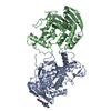
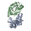
| Title | Crystal structure of a truncated human Tudor-SN | ||||||
 Components Components | Staphylococcal nuclease domain-containing protein 1 | ||||||
 Keywords Keywords |  HYDROLASE / Staphylococcal nuclease OB fold / Tudor domain / Cytoplasm / Host-virus interaction / Nucleus / Phosphoprotein / Transcription / Transcription regulation HYDROLASE / Staphylococcal nuclease OB fold / Tudor domain / Cytoplasm / Host-virus interaction / Nucleus / Phosphoprotein / Transcription / Transcription regulation | ||||||
| Function / homology |  Function and homology information Function and homology informationregulation of cell cycle process / miRNA catabolic process / RISC complex binding / nuclease activity / dense body / regulatory ncRNA-mediated gene silencing / RISC complex / endonuclease activity, active with either ribo- or deoxyribonucleic acids and producing 3'-phosphomonoesters / micrococcal nuclease / mRNA catabolic process ...regulation of cell cycle process / miRNA catabolic process / RISC complex binding / nuclease activity / dense body / regulatory ncRNA-mediated gene silencing / RISC complex / endonuclease activity, active with either ribo- or deoxyribonucleic acids and producing 3'-phosphomonoesters / micrococcal nuclease / mRNA catabolic process / RNA endonuclease activity / transcription coregulator activity / osteoblast differentiation / Signaling by BRAF and RAF1 fusions / melanosome / endonuclease activity / cadherin binding / RNA binding / extracellular exosome / membrane / nucleus / cytosolSimilarity search - Function | ||||||
| Biological species |  Homo sapiens (human) Homo sapiens (human) | ||||||
| Method | X-RAY DIFFRACTION / SYNCHROTRON / MAD / Resolution: 1.9 Å | ||||||
 Authors Authors | Li, C.L. | ||||||
 Citation Citation | Journal: Nucleic Acids Res. / Year: 2008 Title: Structural and functional insights into human Tudor-SN, a key component linking RNA interference and editing. Authors: Li, C.L. / Yang, W.Z. / Chen, Y.P. / Yuan, H.S. | ||||||
| History |
|
- Structure visualization
| Structure viewer | Molecule: MolmilJmol/JSmol |
|---|
- Downloads & links
-Download
| PDBx/mmCIF format | 3bdl.cif.gz | 139 KB | Display | PDBx/mmCIF format |
|---|---|---|---|---|
| PDB format | pdb3bdl.ent.gz | 104.2 KB | Display | PDB format |
| PDBx/mmJSON format | 3bdl.json.gz | Tree view | PDBx/mmJSON format | |
| Others |  Other downloads Other downloads |
-Validation report
| Arichive directory | https://data.pdbj.org/pub/pdb/validation_reports/bd/3bdlftp://data.pdbj.org/pub/pdb/validation_reports/bd/3bdl | HTTPS FTP |
|---|
-Related structure data
| Related structure data | |
|---|---|
| Similar structure data |


-Links
 PDBj
PDBj- Assembly
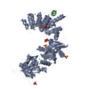
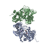
| Deposited unit | 
| ||||||||
|---|---|---|---|---|---|---|---|---|---|
| 1 |
| ||||||||
| 2 | 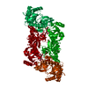
| ||||||||
| Unit cell |
| ||||||||
| Components on special symmetry positions |
|
-Components
| #1: Protein | Mass: 64012.641 Da / Num. of mol.: 1 / Fragment: TSN-64 (SN3, SN4, Tudor, SN5 domains) Source method: isolated from a genetically manipulated source Source: (gene. exp.) Homo sapiens (human) / Gene: SND1, TDRD11 / Plasmid: pET28c / Production host:  Escherichia coli (E. coli) / Strain (production host): B834 / References: UniProt: Q7KZF4, micrococcal nuclease Escherichia coli (E. coli) / Strain (production host): B834 / References: UniProt: Q7KZF4, micrococcal nuclease | ||
|---|---|---|---|
| #2: Chemical | ChemComp-CIT / Citric acid  Mass: 192.124 Da / Num. of mol.: 4 / Source method: obtained synthetically / Formula: C6H8O7 Mass: 192.124 Da / Num. of mol.: 4 / Source method: obtained synthetically / Formula: C6H8O7#3: Water | ChemComp-HOH / | Water Mass: 18.015 Da / Num. of mol.: 690 / Source method: isolated from a natural source / Formula: H2O Mass: 18.015 Da / Num. of mol.: 690 / Source method: isolated from a natural source / Formula: H2O |
-Experimental details
-Experiment
| Experiment | Method: X-RAY DIFFRACTION / Number of used crystals: 1 |
|---|
- Sample preparation
| Crystal | Density Matthews: 3.08 Å3/Da / Density % sol: 60.04 % |
|---|---|
| Crystal grow | Temperature: 283 K / Method: vapor diffusion, hanging drop / pH: 7 Details: 50mM HEPES, pH 7.0, 150mM NaCl, 10mM mercaptoethanol, 1.44M tri-ammonium citrate, VAPOR DIFFUSION, HANGING DROP, temperature 283K |
-Data collection
| Diffraction | Mean temperature: 123 K | ||||||||||||
|---|---|---|---|---|---|---|---|---|---|---|---|---|---|
| Diffraction source | Source: SYNCHROTRON / Site: SPring-8  / Beamline: BL12B2 / Wavelength: 0.979389, 0.979545, 0.964305 / Beamline: BL12B2 / Wavelength: 0.979389, 0.979545, 0.964305 | ||||||||||||
| Detector | Type: ADSC QUANTUM 4 / Detector: CCD / Date: Mar 7, 2007 | ||||||||||||
| Radiation | Monochromator: Fixed-Exit Double Crystal Monochromator / Protocol: MAD / Monochromatic (M) / Laue (L): M / Scattering type: x-ray | ||||||||||||
| Radiation wavelength |
| ||||||||||||
| Reflection | Resolution: 1.82→23.33 Å / Num. all: 68052 / Num. obs: 68052 / % possible obs: 97.8 % / Observed criterion σ(F): 0 / Observed criterion σ(I): 0 / Redundancy: 2.5 % / Biso Wilson estimate: 12.8 Å2 / Rsym value: 0.06 / Net I/σ(I): 15 | ||||||||||||
| Reflection shell | Resolution: 1.82→1.89 Å / Redundancy: 2.5 % / Mean I/σ(I) obs: 2.4 / Num. unique all: 6713 / Rsym value: 0.522 / % possible all: 97.3 |
- Processing
| Software |
| ||||||||||||||||||||||||||||||||||||
|---|---|---|---|---|---|---|---|---|---|---|---|---|---|---|---|---|---|---|---|---|---|---|---|---|---|---|---|---|---|---|---|---|---|---|---|---|---|
| Refinement | Method to determine structure: MAD / Resolution: 1.9→23.33 Å / Rfactor Rfree error: 0.003 / Data cutoff high absF: 405757.73 / Data cutoff low absF: 0 / Isotropic thermal model: RESTRAINED / Cross valid method: THROUGHOUT / σ(F): 0 / σ(I): 0 / Stereochemistry target values: Engh & Huber
| ||||||||||||||||||||||||||||||||||||
| Solvent computation | Solvent model: FLAT MODEL / Bsol: 49.6383 Å2 / ksol: 0.375852 e/Å3 | ||||||||||||||||||||||||||||||||||||
| Displacement parameters | Biso mean: 26.3 Å2
| ||||||||||||||||||||||||||||||||||||
| Refine analyze |
| ||||||||||||||||||||||||||||||||||||
| Refinement step | Cycle: LAST / Resolution: 1.9→23.33 Å
| ||||||||||||||||||||||||||||||||||||
| Refine LS restraints |
| ||||||||||||||||||||||||||||||||||||
| LS refinement shell | Resolution: 1.9→2.02 Å / Rfactor Rfree error: 0.008 / Total num. of bins used: 6
| ||||||||||||||||||||||||||||||||||||
| Xplor file |
|
