 Movie
Movie Controller


+ Open data

- Basic information
| Entry | Database: PDB / ID: 2o4x | ||||||
|---|---|---|---|---|---|---|---|
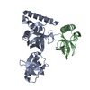
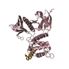
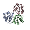
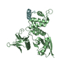
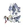
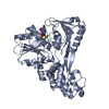
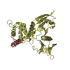
| Title | Crystal structure of human P100 tudor domain | ||||||
 Components Components | (Staphylococcal nuclease domain-containing protein 1) x 2 | ||||||
 Keywords Keywords |  HYDROLASE / OB fold / beta barrel / aromatic cage HYDROLASE / OB fold / beta barrel / aromatic cage | ||||||
| Function / homology |  Function and homology information Function and homology informationregulation of cell cycle process / miRNA catabolic process / RISC complex binding / nuclease activity / dense body / regulatory ncRNA-mediated gene silencing / RISC complex / endonuclease activity, active with either ribo- or deoxyribonucleic acids and producing 3'-phosphomonoesters / micrococcal nuclease / mRNA catabolic process ...regulation of cell cycle process / miRNA catabolic process / RISC complex binding / nuclease activity / dense body / regulatory ncRNA-mediated gene silencing / RISC complex / endonuclease activity, active with either ribo- or deoxyribonucleic acids and producing 3'-phosphomonoesters / micrococcal nuclease / mRNA catabolic process / RNA endonuclease activity / transcription coregulator activity / osteoblast differentiation / Signaling by BRAF and RAF1 fusions / melanosome / endonuclease activity / cadherin binding / RNA binding / extracellular exosome / membrane / nucleus / cytosolSimilarity search - Function | ||||||
| Biological species |  Homo sapiens (human) Homo sapiens (human) | ||||||
| Method | X-RAY DIFFRACTION / SYNCHROTRON / Pt-SAS / Resolution: 2 Å | ||||||
 Authors Authors | Shaw, N. / Zhao, M. / Cheng, C. / Xu, H. / Yang, J. / Silvennoinen, O. / Rao, Z. / Wang, B.C. / Liu, Z.J. | ||||||
 Citation Citation | Journal: To be Published Title: Crystal structure of human P100 tudor domain Authors: Shaw, N. / Zhao, M. / Cheng, C. / Xu, H. / Yang, J. / Silvennoinen, O. / Rao, Z. / Wang, B.C. / Liu, Z.J. | ||||||
| History |
|
- Structure visualization
| Structure viewer | Molecule: MolmilJmol/JSmol |
|---|
- Downloads & links
-Download
| PDBx/mmCIF format | 2o4x.cif.gz | 75.1 KB | Display | PDBx/mmCIF format |
|---|---|---|---|---|
| PDB format | pdb2o4x.ent.gz | 56.7 KB | Display | PDB format |
| PDBx/mmJSON format | 2o4x.json.gz | Tree view | PDBx/mmJSON format | |
| Others |  Other downloads Other downloads |
-Validation report
| Arichive directory | https://data.pdbj.org/pub/pdb/validation_reports/o4/2o4xftp://data.pdbj.org/pub/pdb/validation_reports/o4/2o4x | HTTPS FTP |
|---|
-Related structure data
| Similar structure data |
|---|


-Links
 PDBj
PDBj- Assembly
| Deposited unit | 
| ||||||||
|---|---|---|---|---|---|---|---|---|---|
| 1 |
| ||||||||
| Unit cell |
|
-Components
| #1: Protein | Mass: 24569.662 Da / Num. of mol.: 1 / Fragment: residues 654-870 Source method: isolated from a genetically manipulated source Source: (gene. exp.) Homo sapiens (human) / Plasmid: pGEX6P-1 / Species (production host): Escherichia coli / Production host:  Escherichia coli BL21 (bacteria) / Strain (production host): BL21 / References: UniProt: Q7KZF4 Escherichia coli BL21 (bacteria) / Strain (production host): BL21 / References: UniProt: Q7KZF4 |
|---|---|
| #2: Protein | Mass: 10388.744 Da / Num. of mol.: 1 / Fragment: residues 680-795 Source method: isolated from a genetically manipulated source Source: (gene. exp.) Homo sapiens (human) / Plasmid: pGEX6P-1 / Species (production host): Escherichia coli / Production host: Escherichia coli BL21 (bacteria) / Strain (production host): BL21 / References: UniProt: Q7KZF4 |
| #3: Water | ChemComp-HOH / Water Mass: 18.015 Da / Num. of mol.: 156 / Source method: isolated from a natural source / Formula: H2O Mass: 18.015 Da / Num. of mol.: 156 / Source method: isolated from a natural source / Formula: H2O |
-Experimental details
-Experiment
| Experiment | Method: X-RAY DIFFRACTION / Number of used crystals: 1 |
|---|
- Sample preparation
| Crystal | Density Matthews: 3.18 Å3/Da / Density % sol: 61.28 % |
|---|---|
| Crystal grow | Temperature: 289 K / Method: vapor diffusion, hanging drop / pH: 4.2 Details: 2.0 MICROLITER DROPS CONTAINING EQUAL VOLUMES OF PROTEIN CONCENTRATE (10.0mg/ml), RESERVOIR SOLUTION CONTAING 20% PEG 8000, 100.0mM PHOSPHATE, 0.1M HEPES pH 4.2 , VAPOR DIFFUSION, HANGING DROP, temperature 289K |
-Data collection
| Diffraction | Mean temperature: 100 K |
|---|---|
| Diffraction source | Source: SYNCHROTRON / Site: APS  / Beamline: 22-ID / Wavelength: 0.979 Å / Beamline: 22-ID / Wavelength: 0.979 Å |
| Detector | Type: MARRESEARCH / Detector: CCD / Date: Jun 16, 2006 |
| Radiation | Monochromator: SI Channel 220 / Protocol: SINGLE WAVELENGTH / Monochromatic (M) / Laue (L): M / Scattering type: x-ray |
| Radiation wavelength | Wavelength: 0.979 Å / Relative weight: 1 |
| Reflection | Resolution: 2→44.24 Å / Num. obs: 29102 / Rsym value: 0.078 |
| Reflection shell | Resolution: 2→2.05 Å / Rmerge(I) obs: 0.4 |
- Processing
| Software |
| ||||||||||||||||||||||||||||||||||||||||||||||||||||||||||||||||||||||||||||||||||||||||||
|---|---|---|---|---|---|---|---|---|---|---|---|---|---|---|---|---|---|---|---|---|---|---|---|---|---|---|---|---|---|---|---|---|---|---|---|---|---|---|---|---|---|---|---|---|---|---|---|---|---|---|---|---|---|---|---|---|---|---|---|---|---|---|---|---|---|---|---|---|---|---|---|---|---|---|---|---|---|---|---|---|---|---|---|---|---|---|---|---|---|---|---|
| Refinement | Method to determine structure: Pt-SAS / Resolution: 2→44 Å / Cor.coef. Fo:Fc: 0.929 / Cor.coef. Fo:Fc free: 0.923 / SU B: 3.794 / SU ML: 0.109 / Cross valid method: THROUGHOUT / ESU R: 0.177 / ESU R Free: 0.152 / Stereochemistry target values: MAXIMUM LIKELIHOOD / Details: HYDROGENS HAVE BEEN ADDED IN THE RIDING POSITIONS
| ||||||||||||||||||||||||||||||||||||||||||||||||||||||||||||||||||||||||||||||||||||||||||
| Solvent computation | Ion probe radii: 0.8 Å / Shrinkage radii: 0.8 Å / VDW probe radii: 1.4 Å / Solvent model: MASK | ||||||||||||||||||||||||||||||||||||||||||||||||||||||||||||||||||||||||||||||||||||||||||
| Displacement parameters | Biso mean: 28.325 Å2
| ||||||||||||||||||||||||||||||||||||||||||||||||||||||||||||||||||||||||||||||||||||||||||
| Refinement step | Cycle: LAST / Resolution: 2→44 Å
| ||||||||||||||||||||||||||||||||||||||||||||||||||||||||||||||||||||||||||||||||||||||||||
| Refine LS restraints |
| ||||||||||||||||||||||||||||||||||||||||||||||||||||||||||||||||||||||||||||||||||||||||||
| LS refinement shell | Resolution: 2→2.052 Å / Total num. of bins used: 20
|
