 Movie
Movie Controller

[English] 日本語
 Yorodumi
- PDB-3vp9: Crystal structure of the N-terminal domain of the yeast general c... -
+ Open data

- Basic information
| Entry | Database: PDB / ID: 3vp9 | ||||||
|---|---|---|---|---|---|---|---|
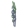
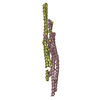
| Title | Crystal structure of the N-terminal domain of the yeast general corepressor Tup1p mutant | ||||||
 Components Components | General transcriptional corepressor TUP1 | ||||||
 Keywords Keywords |  TRANSCRIPTION / Four helix bundle TRANSCRIPTION / Four helix bundle | ||||||
| Function / homology |  Function and homology information Function and homology informationcarbon catabolite repression of transcription from RNA polymerase II promoter by glucose / mediator complex binding / hyperosmotic response / phosphatidylinositol-3,5-bisphosphate binding / transcription repressor complex / histone deacetylase binding / transcription corepressor activity / histone binding / negative regulation of DNA-templated transcription / DNA damage response ...carbon catabolite repression of transcription from RNA polymerase II promoter by glucose / mediator complex binding / hyperosmotic response / phosphatidylinositol-3,5-bisphosphate binding / transcription repressor complex / histone deacetylase binding / transcription corepressor activity / histone binding / negative regulation of DNA-templated transcription / DNA damage response / regulation of transcription by RNA polymerase II / negative regulation of transcription by RNA polymerase II / positive regulation of transcription by RNA polymerase II / nucleusSimilarity search - Function | ||||||
| Biological species |  Saccharomyces cerevisiae (brewer's yeast) Saccharomyces cerevisiae (brewer's yeast) | ||||||
| Method | X-RAY DIFFRACTION / SYNCHROTRON / SAD / Resolution: 1.799 Å | ||||||
 Authors Authors | Matsumura, H. / Kusaka, N. / Nakamura, T. / Tanaka, N. / Sagegami, K. / Uegaki, K. / Inoue, T. / Mukai, Y. | ||||||
 Citation Citation | Journal: J.Biol.Chem. / Year: 2012 Title: Crystal structure of the N-terminal domain of the yeast general corepressor Tup1p and its functional implications Authors: Matsumura, H. / Kusaka, N. / Nakamura, T. / Tanaka, N. / Sagegami, K. / Uegaki, K. / Inoue, T. / Mukai, Y. | ||||||
| History |
|
- Structure visualization
| Structure viewer | Molecule: MolmilJmol/JSmol |
|---|
- Downloads & links
-Download
| PDBx/mmCIF format | 3vp9.cif.gz | 48.3 KB | Display | PDBx/mmCIF format |
|---|---|---|---|---|
| PDB format | pdb3vp9.ent.gz | 35.1 KB | Display | PDB format |
| PDBx/mmJSON format | 3vp9.json.gz | Tree view | PDBx/mmJSON format | |
| Others |  Other downloads Other downloads |
-Validation report
| Arichive directory | https://data.pdbj.org/pub/pdb/validation_reports/vp/3vp9ftp://data.pdbj.org/pub/pdb/validation_reports/vp/3vp9 | HTTPS FTP |
|---|
-Related structure data


-Links
 PDBj
PDBj- Assembly
| Deposited unit | 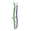
| ||||||||||||||||||
|---|---|---|---|---|---|---|---|---|---|---|---|---|---|---|---|---|---|---|---|
| 1 | 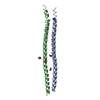
| ||||||||||||||||||
| Unit cell |
| ||||||||||||||||||
| Components on special symmetry positions |
|
-Components
| #1: Protein | Mass: 11086.417 Da / Num. of mol.: 2 / Fragment: N-terminal domain / Mutation: L62R Source method: isolated from a genetically manipulated source Source: (gene. exp.) Saccharomyces cerevisiae (brewer's yeast)Strain: S288c / Gene: TUP1 / Production host:  Escherichia coli (E. coli) / References: UniProt: P16649 Escherichia coli (E. coli) / References: UniProt: P16649#2: Chemical | ChemComp-DIO / 1,4-Dioxane  Mass: 88.105 Da / Num. of mol.: 4 / Source method: obtained synthetically / Formula: C4H8O2 Mass: 88.105 Da / Num. of mol.: 4 / Source method: obtained synthetically / Formula: C4H8O2#3: Water | ChemComp-HOH / | Water Mass: 18.015 Da / Num. of mol.: 174 / Source method: isolated from a natural source / Formula: H2O Mass: 18.015 Da / Num. of mol.: 174 / Source method: isolated from a natural source / Formula: H2O |
|---|
-Experimental details
-Experiment
| Experiment | Method: X-RAY DIFFRACTION / Number of used crystals: 1 |
|---|
- Sample preparation
| Crystal | Density Matthews: 2.71 Å3/Da / Density % sol: 54.66 % |
|---|---|
| Crystal grow | Temperature: 293 K / Method: vapor diffusion / pH: 6.5 Details: 14%(v/v) 1,4-dioxane, 0.1M MES monohydrate, 1.6M ammonium sulfate, pH 6.5, VAPOR DIFFUSION, temperature 293K |
-Data collection
| Diffraction | Mean temperature: 100 K |
|---|---|
| Diffraction source | Source: SYNCHROTRON / Site: Photon Factory  / Beamline: BL-17A / Wavelength: 1 Å / Beamline: BL-17A / Wavelength: 1 Å |
| Detector | Type: ADSC QUANTUM 315 / Detector: CCD / Date: Oct 4, 2010 |
| Radiation | Protocol: SINGLE WAVELENGTH / Monochromatic (M) / Laue (L): M / Scattering type: x-ray |
| Radiation wavelength | Wavelength: 1 Å / Relative weight: 1 |
| Reflection | Resolution: 1.799→409 Å / Num. all: 20399 / Num. obs: 20399 / % possible obs: 91.4 % / Observed criterion σ(F): 2 / Observed criterion σ(I): 2 |
| Reflection shell | Resolution: 1.8→1.83 Å / % possible all: 91.1 |
- Processing
| Software |
| ||||||||||||||||||||||||||||||||||||||||||||||||||||||||
|---|---|---|---|---|---|---|---|---|---|---|---|---|---|---|---|---|---|---|---|---|---|---|---|---|---|---|---|---|---|---|---|---|---|---|---|---|---|---|---|---|---|---|---|---|---|---|---|---|---|---|---|---|---|---|---|---|---|
| Refinement | Method to determine structure: SAD / Resolution: 1.799→33.292 Å / Occupancy max: 1 / Occupancy min: 1 / FOM work R set: 0.8165 / SU ML: 0.14 / σ(F): 0 / Phase error: 25.25 / Stereochemistry target values: ML
| ||||||||||||||||||||||||||||||||||||||||||||||||||||||||
| Solvent computation | Shrinkage radii: 0.72 Å / VDW probe radii: 1 Å / Solvent model: FLAT BULK SOLVENT MODEL / Bsol: 79.794 Å2 / ksol: 0.4 e/Å3 | ||||||||||||||||||||||||||||||||||||||||||||||||||||||||
| Displacement parameters | Biso max: 98.04 Å2 / Biso mean: 45.7701 Å2 / Biso min: 14.38 Å2
| ||||||||||||||||||||||||||||||||||||||||||||||||||||||||
| Refinement step | Cycle: LAST / Resolution: 1.799→33.292 Å
| ||||||||||||||||||||||||||||||||||||||||||||||||||||||||
| Refine LS restraints |
| ||||||||||||||||||||||||||||||||||||||||||||||||||||||||
| LS refinement shell | Refine-ID: X-RAY DIFFRACTION / Total num. of bins used: 7
|
