- PDB-4i0k: Crystal structure of murine B7-H3 extracellular domain -
+
Open data
ID or keywords:
Loading...
-
Basic information
Entry
Database: PDB / ID: 4i0k
Title
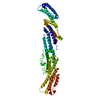
Crystal structure of murine B7-H3 extracellular domain
Components
CD276 antigen
Keywords
IMMUNE SYSTEM / IMMUNOGLOBULIN DOMAIN / GLYCOPROTEIN / IMMUNITY / ADAPTIVE IMMUNITY / Structural Genomics / Protein Structure Initiative / immunoglobulin variable like domain / immunoglobulin constant like domain / cell surface / PSI-Biology / New York Structural Genomics Research Consortium / NYSGRC / Atoms-to-Animals: The Immune Function Network / IFN
Function / homology
Function and homology information
negative regulation of interleukin-2 production / negative regulation of type II interferon production / regulation of immune response / positive regulation of osteoblast differentiation / positive regulation of bone mineralization / regulation of cytokine production / negative regulation of T cell proliferation / positive regulation of T cell proliferation / positive regulation of interleukin-2 production / T cell activation ...negative regulation of interleukin-2 production / negative regulation of type II interferon production / regulation of immune response / positive regulation of osteoblast differentiation / positive regulation of bone mineralization / regulation of cytokine production / negative regulation of T cell proliferation / positive regulation of T cell proliferation / positive regulation of interleukin-2 production / T cell activation / negative regulation of inflammatory response / positive regulation of type II interferon production / T cell receptor signaling pathway / membrane => GO:0016020 / external side of plasma membrane / signaling receptor binding / identical protein binding Similarity search - Function
In the structure databanks used in Yorodumi, some data are registered as the other names, "COVID-19 virus" and "2019-nCoV". Here are the details of the virus and the list of structure data.
Jan 31, 2019. EMDB accession codes are about to change! (news from PDBe EMDB page)
EMDB accession codes are about to change! (news from PDBe EMDB page)
The allocation of 4 digits for EMDB accession codes will soon come to an end. Whilst these codes will remain in use, new EMDB accession codes will include an additional digit and will expand incrementally as the available range of codes is exhausted. The current 4-digit format prefixed with “EMD-” (i.e. EMD-XXXX) will advance to a 5-digit format (i.e. EMD-XXXXX), and so on. It is currently estimated that the 4-digit codes will be depleted around Spring 2019, at which point the 5-digit format will come into force.
The EM Navigator/Yorodumi systems omit the EMD- prefix.
Related info.:Q: What is EMD? / ID/Accession-code notation in Yorodumi/EM Navigator
Yorodumi is a browser for structure data from EMDB, PDB, SASBDB, etc.
This page is also the successor to EM Navigator detail page, and also detail information page/front-end page for Omokage search.
The word "yorodu" (or yorozu) is an old Japanese word meaning "ten thousand". "mi" (miru) is to see.
Related info.:EMDB / PDB / SASBDB / Comparison of 3 databanks / Yorodumi Search / Aug 31, 2016. New EM Navigator & Yorodumi / Yorodumi Papers / Jmol/JSmol / Function and homology information / Changes in new EM Navigator and Yorodumi
 Movie
Movie Controller


 Open data
 Basic information
 Components
 Keywords
 IMMUNE SYSTEM /
  Function and homology information
 Authors
 Citation
 Structure visualization
 Downloads & links
 Other downloads
 PDBj
PDBj


 Assembly


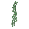
 Type: D-saccharide, beta linking / Mass: 221.208 Da / Num. of mol.: 1
 Mass: 96.063 Da / Num. of mol.: 2 / Source method: obtained synthetically / Formula: SO4
 Mass: 18.015 Da / Num. of mol.: 5 / Source method: isolated from a natural source / Formula: H2O
 Sample preparation
 Processing