 Movie
Movie Controller


+ Open data

- Basic information
| Entry | Database: PDB / ID: 6t68 | ||||||
|---|---|---|---|---|---|---|---|
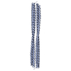
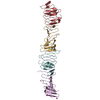
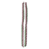
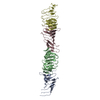
| Title | Crystal structure of Trypanosoma brucei Morn1 | ||||||
 Components Components | MORN repeat-containing protein 1 | ||||||
 Keywords Keywords |  STRUCTURAL PROTEIN / MORN repeat / MORN1 / Trypanosoma STRUCTURAL PROTEIN / MORN repeat / MORN1 / Trypanosoma | ||||||
| Function / homology | bilobe structure / Possible plasma membrane-binding motif in junctophilins, PIP-5-kinases and protein kinases. / MORN motif / MORN repeat / endocytosis / cytosol / MORN repeat-containing protein 1 Function and homology information Function and homology information | ||||||
| Biological species |  Trypanosoma brucei (eukaryote) Trypanosoma brucei (eukaryote) | ||||||
| Method | X-RAY DIFFRACTION / SYNCHROTRON / MOLECULAR REPLACEMENT / Resolution: 2.54 Å | ||||||
 Authors Authors | Grishkovskaya, I. / Kostan, J. / Sajko, S. / Morriswood, B. / Djinovic-Carugo, K. | ||||||
 Citation Citation | Journal: Plos One / Year: 2020 Title: Structures of three MORN repeat proteins and a re-evaluation of the proposed lipid-binding properties of MORN repeats. Authors: Sajko, S. / Grishkovskaya, I. / Kostan, J. / Graewert, M. / Setiawan, K. / Trubestein, L. / Niedermuller, K. / Gehin, C. / Sponga, A. / Puchinger, M. / Gavin, A.C. / Leonard, T.A. / Svergun, ...Authors: Sajko, S. / Grishkovskaya, I. / Kostan, J. / Graewert, M. / Setiawan, K. / Trubestein, L. / Niedermuller, K. / Gehin, C. / Sponga, A. / Puchinger, M. / Gavin, A.C. / Leonard, T.A. / Svergun, D.I. / Smith, T.K. / Morriswood, B. / Djinovic-Carugo, K. | ||||||
| History |
|
- Structure visualization
| Structure viewer | Molecule: MolmilJmol/JSmol |
|---|
- Downloads & links
-Download
| PDBx/mmCIF format | 6t68.cif.gz | 111.4 KB | Display | PDBx/mmCIF format |
|---|---|---|---|---|
| PDB format | pdb6t68.ent.gz | 69.9 KB | Display | PDB format |
| PDBx/mmJSON format | 6t68.json.gz | Tree view | PDBx/mmJSON format | |
| Others |  Other downloads Other downloads |
-Validation report
| Arichive directory | https://data.pdbj.org/pub/pdb/validation_reports/t6/6t68ftp://data.pdbj.org/pub/pdb/validation_reports/t6/6t68 | HTTPS FTP |
|---|
-Related structure data
| Related structure data | 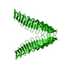 6t4dSC 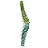 6t4rC 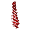 6t69C 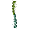 6t6qC S: Starting model for refinement C: citing same article ( |
|---|---|
| Similar structure data |


-Links
 PDBj
PDBj- Assembly
| Deposited unit | 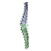
| ||||||||||||
|---|---|---|---|---|---|---|---|---|---|---|---|---|---|
| 1 |
| ||||||||||||
| Unit cell |
|
-Components
| #1: Protein | Mass: 24630.756 Da / Num. of mol.: 2 Source method: isolated from a genetically manipulated source Source: (gene. exp.) Trypanosoma brucei (eukaryote) / Gene: Tb927.6.4670 / Production host:  Escherichia coli (E. coli) / References: UniProt: Q587D3 Escherichia coli (E. coli) / References: UniProt: Q587D3#2: Chemical | ChemComp-CL / Chloride  Mass: 35.453 Da / Num. of mol.: 5 / Source method: obtained synthetically / Formula: Cl Mass: 35.453 Da / Num. of mol.: 5 / Source method: obtained synthetically / Formula: Cl#3: Water | ChemComp-HOH / | Water Mass: 18.015 Da / Num. of mol.: 16 / Source method: isolated from a natural source / Formula: H2O Mass: 18.015 Da / Num. of mol.: 16 / Source method: isolated from a natural source / Formula: H2OHas ligand of interest | N | |
|---|
-Experimental details
-Experiment
| Experiment | Method: X-RAY DIFFRACTION / Number of used crystals: 1 |
|---|
- Sample preparation
| Crystal | Density Matthews: 2.03 Å3/Da / Density % sol: 39.3 % |
|---|---|
| Crystal grow | Temperature: 295.15 K / Method: vapor diffusion, sitting drop Details: 0.166 M Tris-HCl pH 8.8, 0.15 M MgCl2, 0.45 M KI, 24% PEG 2000 MME, and 4% glycerol |
-Data collection
| Diffraction | Mean temperature: 100 K / Serial crystal experiment: N |
|---|---|
| Diffraction source | Source: SYNCHROTRON / Site: ESRF  / Beamline: ID23-1 / Wavelength: 1.89 Å / Beamline: ID23-1 / Wavelength: 1.89 Å |
| Detector | Type: DECTRIS PILATUS 6M / Detector: PIXEL / Date: Dec 2, 2014 |
| Radiation | Protocol: SINGLE WAVELENGTH / Monochromatic (M) / Laue (L): M / Scattering type: x-ray |
| Radiation wavelength | Wavelength: 1.89 Å / Relative weight: 1 |
| Reflection | Resolution: 2.53→48.14 Å / Num. obs: 12419 / % possible obs: 93.1 % / Redundancy: 6.2 % / Biso Wilson estimate: 54 Å2 / CC1/2: 0.995 / Rmerge(I) obs: 0.116 / Rpim(I) all: 0.074 / Net I/σ(I): 9.3 |
| Reflection shell | Resolution: 2.53→2.65 Å / Rmerge(I) obs: 1.365 / Num. unique obs: 975 / CC1/2: 0.303 / % possible all: 60 |
- Processing
| Software |
| |||||||||||||||||||||||||||||||||||
|---|---|---|---|---|---|---|---|---|---|---|---|---|---|---|---|---|---|---|---|---|---|---|---|---|---|---|---|---|---|---|---|---|---|---|---|---|
| Refinement | Method to determine structure: MOLECULAR REPLACEMENT Starting model: 6T4D Resolution: 2.54→48.14 Å / SU ML: 0.4157 / Cross valid method: FREE R-VALUE / σ(F): 1.36 / Phase error: 38.7126
| |||||||||||||||||||||||||||||||||||
| Solvent computation | Shrinkage radii: 0.9 Å / VDW probe radii: 1.11 Å | |||||||||||||||||||||||||||||||||||
| Displacement parameters | Biso mean: 66.4 Å2 | |||||||||||||||||||||||||||||||||||
| Refinement step | Cycle: LAST / Resolution: 2.54→48.14 Å
| |||||||||||||||||||||||||||||||||||
| Refine LS restraints |
| |||||||||||||||||||||||||||||||||||
| LS refinement shell |
|
