 Movie
Movie Controller

[English] 日本語
 Yorodumi
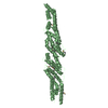
- PDB-4nqi: Structure of the N-terminal I-BAR domain (1-259) of D.Discoideum IBARa -
+ Open data

- Basic information
| Entry | Database: PDB / ID: 4nqi | ||||||
|---|---|---|---|---|---|---|---|
| Title | Structure of the N-terminal I-BAR domain (1-259) of D.Discoideum IBARa | ||||||
 Components Components | SH3 domain-containing protein | ||||||
 Keywords Keywords |  SIGNALING PROTEIN / I-BAR DOMAIN / MEMBRANE REMODELLING / CYTOKINESIS / ENDOCYTOSIS / LIPID BINDING SIGNALING PROTEIN / I-BAR DOMAIN / MEMBRANE REMODELLING / CYTOKINESIS / ENDOCYTOSIS / LIPID BINDING | ||||||
| Function / homology |  Function and homology information Function and homology informationcontractile vacuolar membrane / pore formation during contractile vacuole discharge / sorocarp stalk development / membrane invagination / intracellular water homeostasis / plasma membrane organization / vesicle organization / filopodium tip / cellular hypotonic response / clathrin-dependent endocytosis ...contractile vacuolar membrane / pore formation during contractile vacuole discharge / sorocarp stalk development / membrane invagination / intracellular water homeostasis / plasma membrane organization / vesicle organization / filopodium tip / cellular hypotonic response / clathrin-dependent endocytosis / mitotic cytokinesis / phagocytic vesicle / phagocytosis / actin filament polymerization / cytoskeletal protein binding / phosphatidylinositol binding / microtubule binding / vesicle / cytoskeleton / identical protein binding / plasma membrane / cytoplasmSimilarity search - Function | ||||||
| Biological species |  Dictyostelium discoideum (eukaryote) Dictyostelium discoideum (eukaryote) | ||||||
| Method | X-RAY DIFFRACTION / SYNCHROTRON / SAD / Resolution: 2.21 Å | ||||||
 Authors Authors | Witte, G. / Faix, J. / Runge-Wollmann, P. | ||||||
 Citation Citation | Journal: J.Cell.Sci. / Year: 2014 Title: The inverse BAR domain protein IBARa drives membrane remodeling to control osmoregulation, phagocytosis and cytokinesis. Authors: Linkner, J. / Witte, G. / Zhao, H. / Junemann, A. / Nordholz, B. / Runge-Wollmann, P. / Lappalainen, P. / Faix, J. | ||||||
| History |
|
- Structure visualization
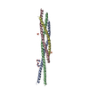
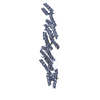
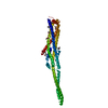
| Structure viewer | Molecule: MolmilJmol/JSmol |
|---|
- Downloads & links
-Download
| PDBx/mmCIF format | 4nqi.cif.gz | 371.9 KB | Display | PDBx/mmCIF format |
|---|---|---|---|---|
| PDB format | pdb4nqi.ent.gz | 316.3 KB | Display | PDB format |
| PDBx/mmJSON format | 4nqi.json.gz | Tree view | PDBx/mmJSON format | |
| Others |  Other downloads Other downloads |
-Validation report
| Arichive directory | https://data.pdbj.org/pub/pdb/validation_reports/nq/4nqiftp://data.pdbj.org/pub/pdb/validation_reports/nq/4nqi | HTTPS FTP |
|---|
-Related structure data
| Similar structure data |
|---|

-Links
 PDBj
PDBj
- Assembly
| Deposited unit | 
| ||||||||
|---|---|---|---|---|---|---|---|---|---|
| 1 | 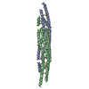
| ||||||||
| 2 | 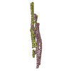
| ||||||||
| Unit cell |
|
-Components
| #1: Protein | Mass: 30242.424 Da / Num. of mol.: 4 Source method: isolated from a genetically manipulated source Source: (gene. exp.) Dictyostelium discoideum (eukaryote) / Gene: DDB_G0274805 / Production host:  Escherichia coli (E. coli) / References: UniProt: C7FZZ0 Escherichia coli (E. coli) / References: UniProt: C7FZZ0#2: Chemical | Diethylene glycol  Mass: 106.120 Da / Num. of mol.: 3 / Source method: obtained synthetically / Formula: C4H10O3 Mass: 106.120 Da / Num. of mol.: 3 / Source method: obtained synthetically / Formula: C4H10O3#3: Chemical | Acetate  Mass: 59.044 Da / Num. of mol.: 2 / Source method: obtained synthetically / Formula: C2H3O2 Mass: 59.044 Da / Num. of mol.: 2 / Source method: obtained synthetically / Formula: C2H3O2#4: Water | ChemComp-HOH / | Water Mass: 18.015 Da / Num. of mol.: 276 / Source method: isolated from a natural source / Formula: H2O Mass: 18.015 Da / Num. of mol.: 276 / Source method: isolated from a natural source / Formula: H2OSequence details | AUTHORS HAVE AMPLIFIED THE GENE FROM GENOMIC DNA OF D.DISCOIDEUM AX2 LABORATORY STRAIN AND THE ...AUTHORS HAVE AMPLIFIED THE GENE FROM GENOMIC DNA OF D.DISCOIDEUM | |
|---|
-Experimental details
-Experiment
| Experiment | Method: X-RAY DIFFRACTION / Number of used crystals: 1 |
|---|
- Sample preparation
| Crystal | Density Matthews: 2.14 Å3/Da / Density % sol: 42.41 % |
|---|---|
| Crystal grow | Temperature: 293 K / Method: vapor diffusion, hanging drop / pH: 7 Details: 20 % PEG 3350, 0.25M ammonium acetate, 15% v/v glycerol, pH 7, VAPOR DIFFUSION, HANGING DROP, temperature 293K |
-Data collection
| Diffraction | Mean temperature: 100 K |
|---|---|
| Diffraction source | Source: SYNCHROTRON / Site: SLS  / Beamline: X06SA / Wavelength: 0.9796 Å / Beamline: X06SA / Wavelength: 0.9796 Å |
| Detector | Type: MARMOSAIC 225 mm CCD / Detector: CCD |
| Radiation | Monochromator: Si 111 / Protocol: SINGLE WAVELENGTH / Monochromatic (M) / Laue (L): M / Scattering type: x-ray |
| Radiation wavelength | Wavelength: 0.9796 Å / Relative weight: 1 |
| Reflection | Resolution: 2.21→50 Å / Num. all: 100735 / Num. obs: 99218 / % possible obs: 98.5 % / Observed criterion σ(I): -3 / Net I/σ(I): 10.21 |
| Reflection shell | Resolution: 2.21→2.26 Å / Redundancy: 2.48 % / Mean I/σ(I) obs: 1.88 / Num. unique all: 7425 / % possible all: 85.3 |
- Processing
| Software |
| |||||||||||||||||||||||||||||||||||||||||||||||||||||||||||||||||||||||||||||||||||||||||||||||||||||||||||||||||||||||||||||||||||||||||||||||||||||||||||||||||||||||||||||||||||||||||||||||||||||||||||||||||||||||||
|---|---|---|---|---|---|---|---|---|---|---|---|---|---|---|---|---|---|---|---|---|---|---|---|---|---|---|---|---|---|---|---|---|---|---|---|---|---|---|---|---|---|---|---|---|---|---|---|---|---|---|---|---|---|---|---|---|---|---|---|---|---|---|---|---|---|---|---|---|---|---|---|---|---|---|---|---|---|---|---|---|---|---|---|---|---|---|---|---|---|---|---|---|---|---|---|---|---|---|---|---|---|---|---|---|---|---|---|---|---|---|---|---|---|---|---|---|---|---|---|---|---|---|---|---|---|---|---|---|---|---|---|---|---|---|---|---|---|---|---|---|---|---|---|---|---|---|---|---|---|---|---|---|---|---|---|---|---|---|---|---|---|---|---|---|---|---|---|---|---|---|---|---|---|---|---|---|---|---|---|---|---|---|---|---|---|---|---|---|---|---|---|---|---|---|---|---|---|---|---|---|---|---|---|---|---|---|---|---|---|---|---|---|---|---|---|---|---|---|
| Refinement | Method to determine structure: SAD / Resolution: 2.21→48.401 Å / SU ML: 0.26 / σ(F): 0.88 / Phase error: 24.92 / Stereochemistry target values: ML
| |||||||||||||||||||||||||||||||||||||||||||||||||||||||||||||||||||||||||||||||||||||||||||||||||||||||||||||||||||||||||||||||||||||||||||||||||||||||||||||||||||||||||||||||||||||||||||||||||||||||||||||||||||||||||
| Solvent computation | Shrinkage radii: 0.9 Å / VDW probe radii: 1.11 Å / Solvent model: FLAT BULK SOLVENT MODEL | |||||||||||||||||||||||||||||||||||||||||||||||||||||||||||||||||||||||||||||||||||||||||||||||||||||||||||||||||||||||||||||||||||||||||||||||||||||||||||||||||||||||||||||||||||||||||||||||||||||||||||||||||||||||||
| Refinement step | Cycle: LAST / Resolution: 2.21→48.401 Å
| |||||||||||||||||||||||||||||||||||||||||||||||||||||||||||||||||||||||||||||||||||||||||||||||||||||||||||||||||||||||||||||||||||||||||||||||||||||||||||||||||||||||||||||||||||||||||||||||||||||||||||||||||||||||||
| Refine LS restraints |
| |||||||||||||||||||||||||||||||||||||||||||||||||||||||||||||||||||||||||||||||||||||||||||||||||||||||||||||||||||||||||||||||||||||||||||||||||||||||||||||||||||||||||||||||||||||||||||||||||||||||||||||||||||||||||
| LS refinement shell |
|
