 Movie
Movie Controller


+ Open data

- Basic information
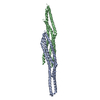
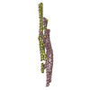
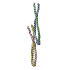
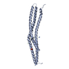
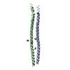
| Entry | Database: PDB / ID: 1y2o | ||||||
|---|---|---|---|---|---|---|---|
| Title | Structure of N-terminal domain IRSp53/BAIAP2 | ||||||
 Components Components | BAI1-associated protein 2 isoform 1 | ||||||
 Keywords Keywords |  SIGNALING PROTEIN / cell motility / filopodia / actin bundling SIGNALING PROTEIN / cell motility / filopodia / actin bundling | ||||||
| Function / homology |  Function and homology information Function and homology informationneuron projection branch point / dendritic spine cytoplasm / plasma membrane organization / actin crosslink formation / positive regulation of dendritic spine morphogenesis / cellular response to L-glutamate / protein localization to synapse / cadherin binding involved in cell-cell adhesion / cytoskeletal anchor activity / regulation of modification of postsynaptic actin cytoskeleton ...neuron projection branch point / dendritic spine cytoplasm / plasma membrane organization / actin crosslink formation / positive regulation of dendritic spine morphogenesis / cellular response to L-glutamate / protein localization to synapse / cadherin binding involved in cell-cell adhesion / cytoskeletal anchor activity / regulation of modification of postsynaptic actin cytoskeleton / presynaptic cytosol / neuron projection terminus / proline-rich region binding / postsynaptic cytosol / positive regulation of actin filament polymerization / dendrite development / positive regulation of excitatory postsynaptic potential / actin filament bundle assembly / CDC42 GTPase cycle / excitatory synapse / RHO GTPases Activate WASPs and WAVEs / RAC3 GTPase cycle / postsynaptic density, intracellular component / cellular response to epidermal growth factor stimulus / ruffle / RAC1 GTPase cycle / axonogenesis / dendritic shaft / secretory granule / synaptic membrane / filopodium / PDZ domain binding / transcription coregulator binding / regulation of actin cytoskeleton organization / FCGR3A-mediated phagocytosis / adherens junction / Schaffer collateral - CA1 synapse / regulation of synaptic plasticity / Regulation of actin dynamics for phagocytic cup formation / VEGFA-VEGFR2 Pathway / insulin receptor signaling pathway / lamellipodium / regulation of cell shape / scaffold protein binding / microtubule / neuronal cell body / glutamatergic synapse / extracellular exosome / nucleoplasm / identical protein binding / plasma membrane / cytosol / cytoplasmSimilarity search - Function | ||||||
| Biological species |  Homo sapiens (human) Homo sapiens (human) | ||||||
| Method | X-RAY DIFFRACTION / SYNCHROTRON / MAD / Resolution: 2.2 Å | ||||||
 Authors Authors | Millard, T.H. / Bompard, G. / Heung, M.-Y. / Dafforn, T.R. / Scott, D.J. / Machesky, L.M. / Futterer, K. | ||||||
 Citation Citation | Journal: Embo J. / Year: 2005 Title: Structural basis of filopodia formation induced by the IRSp53/MIM homology domain of human IRSp53 Authors: Millard, T.H. / Bompard, G. / Heung, M.-Y. / Dafforn, T.R. / Scott, D.J. / Machesky, L.M. / Futterer, K. | ||||||
| History |
|
- Structure visualization
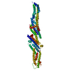
| Structure viewer | Molecule: MolmilJmol/JSmol |
|---|
- Downloads & links
-Download
| PDBx/mmCIF format | 1y2o.cif.gz | 108.3 KB | Display | PDBx/mmCIF format |
|---|---|---|---|---|
| PDB format | pdb1y2o.ent.gz | 89.8 KB | Display | PDB format |
| PDBx/mmJSON format | 1y2o.json.gz | Tree view | PDBx/mmJSON format | |
| Others |  Other downloads Other downloads |
-Validation report
| Arichive directory | https://data.pdbj.org/pub/pdb/validation_reports/y2/1y2oftp://data.pdbj.org/pub/pdb/validation_reports/y2/1y2o | HTTPS FTP |
|---|
-Related structure data
| Similar structure data |
|---|


-Links
 PDBj
PDBj


- Assembly
| Deposited unit | 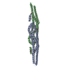
| ||||||||
|---|---|---|---|---|---|---|---|---|---|
| 1 |
| ||||||||
| Unit cell |
|
-Components
| #1: Protein | Mass: 28979.770 Da / Num. of mol.: 2 / Fragment: N-terminal domain Source method: isolated from a genetically manipulated source Source: (gene. exp.) Homo sapiens (human) / Plasmid: pGEX2T / Production host:  Escherichia coli (E. coli) / Strain (production host): B834(DE3) / References: UniProt: Q9UQB8 Escherichia coli (E. coli) / Strain (production host): B834(DE3) / References: UniProt: Q9UQB8#2: Water | ChemComp-HOH / | Water Mass: 18.015 Da / Num. of mol.: 221 / Source method: isolated from a natural source / Formula: H2O Mass: 18.015 Da / Num. of mol.: 221 / Source method: isolated from a natural source / Formula: H2O |
|---|
-Experimental details
-Experiment
| Experiment | Method: X-RAY DIFFRACTION / Number of used crystals: 1 |
|---|
- Sample preparation
| Crystal | Density Matthews: 1.92 Å3/Da / Density % sol: 33.5 % |
|---|
-Data collection
| Diffraction | Mean temperature: 100 K | ||||||||||||
|---|---|---|---|---|---|---|---|---|---|---|---|---|---|
| Diffraction source | Source: SYNCHROTRON / Site: SRS  / Beamline: PX14.2 / Wavelength: 0.9781, 0.9776, 0.9649 / Beamline: PX14.2 / Wavelength: 0.9781, 0.9776, 0.9649 | ||||||||||||
| Detector | Type: ADSC QUANTUM 4 / Detector: CCD / Date: Jul 25, 2004 | ||||||||||||
| Radiation | Protocol: MAD / Monochromatic (M) / Laue (L): M / Scattering type: x-ray | ||||||||||||
| Radiation wavelength |
| ||||||||||||
| Reflection | Resolution: 2.2→47.8 Å / Num. all: 22459 / Num. obs: 22459 / % possible obs: 97.4 % / Observed criterion σ(F): 0 / Observed criterion σ(I): 0 / Redundancy: 3.9 % / Biso Wilson estimate: 35.3 Å2 / Rmerge(I) obs: 0.056 / Rsym value: 0.056 / Net I/σ(I): 25.9 | ||||||||||||
| Reflection shell | Resolution: 2.2→2.32 Å / Redundancy: 3.7 % / Rmerge(I) obs: 0.192 / Mean I/σ(I) obs: 8.4 / Num. unique all: 3133 / Rsym value: 0.192 / % possible all: 93.7 |
- Processing
| Software |
| ||||||||||||||||||||||||||||||||||||||||||||||||||||||||||||||||||||||||||||||||||||||||||
|---|---|---|---|---|---|---|---|---|---|---|---|---|---|---|---|---|---|---|---|---|---|---|---|---|---|---|---|---|---|---|---|---|---|---|---|---|---|---|---|---|---|---|---|---|---|---|---|---|---|---|---|---|---|---|---|---|---|---|---|---|---|---|---|---|---|---|---|---|---|---|---|---|---|---|---|---|---|---|---|---|---|---|---|---|---|---|---|---|---|---|---|
| Refinement | Method to determine structure: MAD / Resolution: 2.2→47.8 Å / Cor.coef. Fo:Fc: 0.929 / Cor.coef. Fo:Fc free: 0.901 / SU B: 14.899 / SU ML: 0.192 / Cross valid method: THROUGHOUT / σ(F): 0 / ESU R: 0.493 / ESU R Free: 0.267 / Stereochemistry target values: MAXIMUM LIKELIHOOD
| ||||||||||||||||||||||||||||||||||||||||||||||||||||||||||||||||||||||||||||||||||||||||||
| Solvent computation | Ion probe radii: 0.8 Å / Shrinkage radii: 0.8 Å / VDW probe radii: 1.2 Å / Solvent model: BABINET MODEL WITH MASK | ||||||||||||||||||||||||||||||||||||||||||||||||||||||||||||||||||||||||||||||||||||||||||
| Displacement parameters | Biso mean: 36.328 Å2
| ||||||||||||||||||||||||||||||||||||||||||||||||||||||||||||||||||||||||||||||||||||||||||
| Refinement step | Cycle: LAST / Resolution: 2.2→47.8 Å
| ||||||||||||||||||||||||||||||||||||||||||||||||||||||||||||||||||||||||||||||||||||||||||
| Refine LS restraints |
| ||||||||||||||||||||||||||||||||||||||||||||||||||||||||||||||||||||||||||||||||||||||||||
| LS refinement shell | Resolution: 2.2→2.319 Å / Total num. of bins used: 10
| ||||||||||||||||||||||||||||||||||||||||||||||||||||||||||||||||||||||||||||||||||||||||||
| Refinement TLS params. | Method: refined / Refine-ID: X-RAY DIFFRACTION
| ||||||||||||||||||||||||||||||||||||||||||||||||||||||||||||||||||||||||||||||||||||||||||
| Refinement TLS group |
|
