 Movie
Movie Controller


+ Open data

- Basic information
| Entry | Database: PDB / ID: 1wdz | ||||||
|---|---|---|---|---|---|---|---|
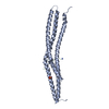
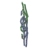
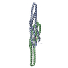
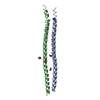
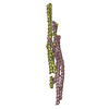
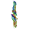
| Title | Crystal structure of RCB domain of IRSp53 | ||||||
 Components Components | insulin receptor substrate p53 | ||||||
 Keywords Keywords |  SIGNALING PROTEIN / cellular signaling protein / RIKEN Structural Genomics/Proteomics Initiative / RSGI / Structural Genomics SIGNALING PROTEIN / cellular signaling protein / RIKEN Structural Genomics/Proteomics Initiative / RSGI / Structural Genomics | ||||||
| Function / homology |  Function and homology information Function and homology informationneuron projection branch point / dendritic spine cytoplasm / plasma membrane organization / actin crosslink formation / positive regulation of dendritic spine morphogenesis / cellular response to L-glutamate / protein localization to synapse / cadherin binding involved in cell-cell adhesion / cytoskeletal anchor activity / regulation of modification of postsynaptic actin cytoskeleton ...neuron projection branch point / dendritic spine cytoplasm / plasma membrane organization / actin crosslink formation / positive regulation of dendritic spine morphogenesis / cellular response to L-glutamate / protein localization to synapse / cadherin binding involved in cell-cell adhesion / cytoskeletal anchor activity / regulation of modification of postsynaptic actin cytoskeleton / presynaptic cytosol / neuron projection terminus / proline-rich region binding / postsynaptic cytosol / positive regulation of actin filament polymerization / dendrite development / positive regulation of excitatory postsynaptic potential / actin filament bundle assembly / CDC42 GTPase cycle / excitatory synapse / RHO GTPases Activate WASPs and WAVEs / RAC3 GTPase cycle / postsynaptic density, intracellular component / cellular response to epidermal growth factor stimulus / ruffle / RAC1 GTPase cycle / axonogenesis / dendritic shaft / secretory granule / synaptic membrane / filopodium / PDZ domain binding / transcription coregulator binding / regulation of actin cytoskeleton organization / FCGR3A-mediated phagocytosis / adherens junction / Schaffer collateral - CA1 synapse / regulation of synaptic plasticity / Regulation of actin dynamics for phagocytic cup formation / VEGFA-VEGFR2 Pathway / insulin receptor signaling pathway / lamellipodium / regulation of cell shape / scaffold protein binding / microtubule / neuronal cell body / glutamatergic synapse / extracellular exosome / nucleoplasm / identical protein binding / plasma membrane / cytosol / cytoplasmSimilarity search - Function | ||||||
| Biological species |  Homo sapiens (human) Homo sapiens (human) | ||||||
| Method | X-RAY DIFFRACTION / SYNCHROTRON / SIRAS / Resolution: 2.63 Å | ||||||
 Authors Authors | Murayama, K. / Suetsugu, S. / Seto, A. / Shirouzu, M. / Takenawa, T. / Yokoyama, S. / RIKEN Structural Genomics/Proteomics Initiative (RSGI) | ||||||
 Citation Citation | Journal: TO BE PUBLISHED Title: Crystal structure of RCB domain of IRSp53 Authors: Murayama, K. / Suetsugu, S. / Seto, A. / Shirouzu, M. / Takenawa, T. / Yokoyama, S. | ||||||
| History |
|
- Structure visualization
| Structure viewer | Molecule: MolmilJmol/JSmol |
|---|
- Downloads & links
-Download
| PDBx/mmCIF format | 1wdz.cif.gz | 100.3 KB | Display | PDBx/mmCIF format |
|---|---|---|---|---|
| PDB format | pdb1wdz.ent.gz | 78.2 KB | Display | PDB format |
| PDBx/mmJSON format | 1wdz.json.gz | Tree view | PDBx/mmJSON format | |
| Others |  Other downloads Other downloads |
-Validation report
| Arichive directory | https://data.pdbj.org/pub/pdb/validation_reports/wd/1wdzftp://data.pdbj.org/pub/pdb/validation_reports/wd/1wdz | HTTPS FTP |
|---|
-Related structure data
| Similar structure data | |
|---|---|
| Other databases |


-Links
 PDBj
PDBj


- Assembly
| Deposited unit | 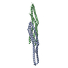
| ||||||||
|---|---|---|---|---|---|---|---|---|---|
| 1 |
| ||||||||
| Unit cell |
|
-Components
| #1: Protein | Mass: 27597.312 Da / Num. of mol.: 2 / Fragment: N-terminal domain Source method: isolated from a genetically manipulated source Source: (gene. exp.) Homo sapiens (human) / Plasmid: pGEX-4T-1 / Production host:  Escherichia coli (E. coli) / References: UniProt: Q9UQB8 Escherichia coli (E. coli) / References: UniProt: Q9UQB8#2: Water | ChemComp-HOH / | Water Mass: 18.015 Da / Num. of mol.: 70 / Source method: isolated from a natural source / Formula: H2O Mass: 18.015 Da / Num. of mol.: 70 / Source method: isolated from a natural source / Formula: H2O |
|---|
-Experimental details
-Experiment
| Experiment | Method: X-RAY DIFFRACTION / Number of used crystals: 1 |
|---|
- Sample preparation
| Crystal | Density Matthews: 2.34 Å3/Da / Density % sol: 47.4 % |
|---|---|
| Crystal grow | Temperature: 293 K / Method: vapor diffusion, hanging drop / pH: 8.5 Details: 27% PEG4000, 0.09M Tris pH8.5, 0.19M Sodium acetate, 3.5% MPD, VAPOR DIFFUSION, HANGING DROP, temperature 293K |
-Data collection
| Diffraction | Mean temperature: 100 K |
|---|---|
| Diffraction source | Source: SYNCHROTRON / Site: SPring-8  / Beamline: BL44B2 / Wavelength: 1 Å / Beamline: BL44B2 / Wavelength: 1 Å |
| Detector | Type: MARRESEARCH / Detector: CCD / Date: Oct 22, 2002 |
| Radiation | Monochromator: Si 111 CHANNEL / Protocol: SINGLE WAVELENGTH / Monochromatic (M) / Laue (L): M / Scattering type: x-ray |
| Radiation wavelength | Wavelength: 1 Å / Relative weight: 1 |
| Reflection | Resolution: 2.63→50 Å / Num. obs: 15858 / % possible obs: 98.6 % / Observed criterion σ(I): -3 / Redundancy: 4.2 % / Biso Wilson estimate: 38.4 Å2 / Rsym value: 0.059 / Net I/σ(I): 26.1 |
| Reflection shell | Resolution: 2.63→2.75 Å / Mean I/σ(I) obs: 5.5 / Rsym value: 0.303 / % possible all: 98.2 |
- Processing
| Software |
| ||||||||||||||||||||
|---|---|---|---|---|---|---|---|---|---|---|---|---|---|---|---|---|---|---|---|---|---|
| Refinement | Method to determine structure: SIRAS / Resolution: 2.63→41.94 Å / Rfactor Rfree error: 0.007 / Data cutoff high absF: 576339.67 / Data cutoff low absF: 0 / Isotropic thermal model: RESTRAINED / Cross valid method: THROUGHOUT / σ(F): 0
| ||||||||||||||||||||
| Solvent computation | Solvent model: FLAT MODEL / Bsol: 25.4682 Å2 / ksol: 0.293805 e/Å3 | ||||||||||||||||||||
| Displacement parameters | Biso mean: 51 Å2
| ||||||||||||||||||||
| Refine analyze |
| ||||||||||||||||||||
| Refinement step | Cycle: LAST / Resolution: 2.63→41.94 Å
| ||||||||||||||||||||
| Refine LS restraints |
| ||||||||||||||||||||
| LS refinement shell | Resolution: 2.63→2.79 Å / Rfactor Rfree error: 0.025 / Total num. of bins used: 6
| ||||||||||||||||||||
| Xplor file |
|
