 Movie
Movie Controller

[English] 日本語
 Yorodumi
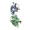
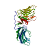
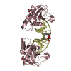
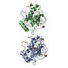
- PDB-3t5g: Structure of fully modified farnesylated Rheb in complex with PDE6D -
+ Open data

- Basic information
| Entry | Database: PDB / ID: 3t5g | ||||||
|---|---|---|---|---|---|---|---|
| Title | Structure of fully modified farnesylated Rheb in complex with PDE6D | ||||||
 Components Components |
| ||||||
 Keywords Keywords |  SIGNALING PROTEIN / LIPID BINDING PROTEIN / Immunoglobulin-like beta sandwitch / PDE delta / Rheb / Farnesyl SIGNALING PROTEIN / LIPID BINDING PROTEIN / Immunoglobulin-like beta sandwitch / PDE delta / Rheb / Farnesyl | ||||||
| Function / homology |  Function and homology information Function and homology informationARL13B-mediated ciliary trafficking of INPP5E / regulation of type B pancreatic cell development / GTPase inhibitor activity / MTOR signalling / Amino acids regulate mTORC1 / Energy dependent regulation of mTOR by LKB1-AMPK / negative regulation of cold-induced thermogenesis / response to stimulus / Macroautophagy / small GTPase-mediated signal transduction ...ARL13B-mediated ciliary trafficking of INPP5E / regulation of type B pancreatic cell development / GTPase inhibitor activity / MTOR signalling / Amino acids regulate mTORC1 / Energy dependent regulation of mTOR by LKB1-AMPK / negative regulation of cold-induced thermogenesis / response to stimulus / Macroautophagy / small GTPase-mediated signal transduction / positive regulation of oligodendrocyte differentiation / protein kinase activator activity / oligodendrocyte differentiation / mTORC1-mediated signalling / cellular response to nutrient levels / positive regulation of TOR signaling / regulation of macroautophagy / endomembrane system / positive regulation of TORC1 signaling / visual perception / protein serine/threonine kinase activator activity / Regulation of PTEN gene transcription / TP53 Regulates Metabolic Genes / Hydrolases; Acting on acid anhydrides; Acting on GTP to facilitate cellular and subcellular movement / spliceosomal complex / cytoplasmic vesicle membrane / cilium / small GTPase binding / RAS processing / GDP binding / cytoplasmic vesicle / postsynaptic density / cytoskeleton / regulation of cell cycle / lysosomal membrane / Golgi membrane / GTPase activity / endoplasmic reticulum membrane / GTP binding / protein kinase binding / magnesium ion binding / signal transduction / extracellular exosome / membrane / plasma membrane / cytosol / cytoplasmSimilarity search - Function | ||||||
| Biological species |  Homo sapiens (human) Homo sapiens (human) | ||||||
| Method | X-RAY DIFFRACTION / SYNCHROTRON / MOLECULAR REPLACEMENT / Resolution: 1.7 Å | ||||||
 Authors Authors | Ismail, S.A. / Chen, Y.-X. / Wittinghofer, A. | ||||||
 Citation Citation | Journal: Nat.Chem.Biol. / Year: 2011 Title: Arl2-GTP and Arl3-GTP regulate a GDI-like transport system for farnesylated cargo. Authors: Ismail, S.A. / Chen, Y.X. / Rusinova, A. / Chandra, A. / Bierbaum, M. / Gremer, L. / Triola, G. / Waldmann, H. / Bastiaens, P.I. / Wittinghofer, A. | ||||||
| History |
|
- Structure visualization
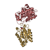
| Structure viewer | Molecule: MolmilJmol/JSmol |
|---|
- Downloads & links
-Download
| PDBx/mmCIF format | 3t5g.cif.gz | 91.1 KB | Display | PDBx/mmCIF format |
|---|---|---|---|---|
| PDB format | pdb3t5g.ent.gz | 65.8 KB | Display | PDB format |
| PDBx/mmJSON format | 3t5g.json.gz | Tree view | PDBx/mmJSON format | |
| Others |  Other downloads Other downloads |
-Validation report
| Arichive directory | https://data.pdbj.org/pub/pdb/validation_reports/t5/3t5gftp://data.pdbj.org/pub/pdb/validation_reports/t5/3t5g | HTTPS FTP |
|---|
-Related structure data
| Related structure data | 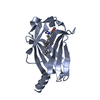 3t5iC 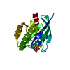 1xtqS C: citing same article ( S: Starting model for refinement |
|---|---|
| Similar structure data |


-Links
 PDBj
PDBj


- Assembly
| Deposited unit | 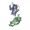
| ||||||||
|---|---|---|---|---|---|---|---|---|---|
| 1 |
| ||||||||
| Unit cell |
|
-Components
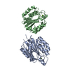
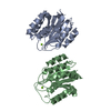
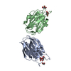
-Protein , 2 types, 2 molecules AB
| #1: Protein | Mass: 20248.135 Da / Num. of mol.: 1 Source method: isolated from a genetically manipulated source Source: (gene. exp.) Homo sapiens (human) / Gene: RHEB, RHEB2 / Production host:  Escherichia coli (E. coli) / References: UniProt: Q15382 Escherichia coli (E. coli) / References: UniProt: Q15382 |
|---|---|
| #2: Protein | Mass: 17585.121 Da / Num. of mol.: 1 Source method: isolated from a genetically manipulated source Source: (gene. exp.) Homo sapiens (human) / Gene: PDE6D, PDED / Production host: Escherichia coli (E. coli) / References: UniProt: O43924 |
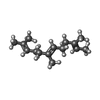
-Non-polymers , 4 types, 419 molecules 


| #3: Chemical | ChemComp-GDP / Guanosine diphosphate Type: RNA linking / Mass: 443.201 Da / Num. of mol.: 1 / Source method: obtained synthetically / Formula: C10H15N5O11P2 / Comment: GDP, energy-carrying molecule*YM Type: RNA linking / Mass: 443.201 Da / Num. of mol.: 1 / Source method: obtained synthetically / Formula: C10H15N5O11P2 / Comment: GDP, energy-carrying molecule*YM |
|---|---|
| #4: Chemical | ChemComp-MG /  Mass: 24.305 Da / Num. of mol.: 1 / Source method: obtained synthetically / Formula: Mg Mass: 24.305 Da / Num. of mol.: 1 / Source method: obtained synthetically / Formula: Mg |
| #5: Chemical | ChemComp-FAR / Farnesol Mass: 206.367 Da / Num. of mol.: 1 / Source method: obtained synthetically / Formula: C15H26 Mass: 206.367 Da / Num. of mol.: 1 / Source method: obtained synthetically / Formula: C15H26 |
| #6: Water | ChemComp-HOH / WaterMass: 18.015 Da / Num. of mol.: 416 / Source method: isolated from a natural source / Formula: H2O |
-Experimental details
-Experiment
| Experiment | Method: X-RAY DIFFRACTION / Number of used crystals: 1 |
|---|
- Sample preparation
| Crystal | Density Matthews: 2.44 Å3/Da / Density % sol: 49.64 % |
|---|---|
| Crystal grow | Temperature: 293 K / Method: vapor diffusion, hanging drop / pH: 7.2 Details: 0.1 M Tris 8.5, and 12.5 % PEG8000, pH 7.2, VAPOR DIFFUSION, HANGING DROP, temperature 293K |
-Data collection
| Diffraction | Mean temperature: 100 K |
|---|---|
| Diffraction source | Source: SYNCHROTRON / Site: SLS  / Beamline: X10SA / Wavelength: 0.979 Å / Beamline: X10SA / Wavelength: 0.979 Å |
| Detector | Type: MARMOSAIC 225 mm CCD / Detector: CCD / Date: Jul 31, 2010 |
| Radiation | Monochromator: SAGITALLY - HORIZONTALLY FOCUSED SI(111) MONOCHROMATOR Protocol: SINGLE WAVELENGTH / Monochromatic (M) / Laue (L): M / Scattering type: x-ray |
| Radiation wavelength | Wavelength: 0.979 Å / Relative weight: 1 |
| Reflection | Resolution: 1.7→30 Å / Num. all: 40454 / Num. obs: 40454 / % possible obs: 97.2 % / Observed criterion σ(F): 1 / Observed criterion σ(I): 1 |
| Reflection shell | Resolution: 1.7→1.8 Å / Redundancy: 6.7 % / Rmerge(I) obs: 0.28 / Mean I/σ(I) obs: 5.95 / Num. unique all: 6158 / % possible all: 95.7 |
- Processing
| Software |
| |||||||||||||||||||||||||||||||||||||||||||||||||||||||||||||||||
|---|---|---|---|---|---|---|---|---|---|---|---|---|---|---|---|---|---|---|---|---|---|---|---|---|---|---|---|---|---|---|---|---|---|---|---|---|---|---|---|---|---|---|---|---|---|---|---|---|---|---|---|---|---|---|---|---|---|---|---|---|---|---|---|---|---|---|
| Refinement | Method to determine structure: MOLECULAR REPLACEMENT Starting model: 1XTQ Resolution: 1.7→29.01 Å / Cor.coef. Fo:Fc: 0.95 / Cor.coef. Fo:Fc free: 0.932 / SU B: 2.211 / SU ML: 0.074 / Cross valid method: THROUGHOUT / σ(F): 1 / ESU R Free: 0.114 / Stereochemistry target values: MAXIMUM LIKELIHOOD / Details: HYDROGENS HAVE BEEN ADDED IN THE RIDING POSITIONS
| |||||||||||||||||||||||||||||||||||||||||||||||||||||||||||||||||
| Solvent computation | Ion probe radii: 0.8 Å / Shrinkage radii: 0.8 Å / VDW probe radii: 1.4 Å / Solvent model: MASK | |||||||||||||||||||||||||||||||||||||||||||||||||||||||||||||||||
| Displacement parameters | Biso mean: 19.637 Å2
| |||||||||||||||||||||||||||||||||||||||||||||||||||||||||||||||||
| Refinement step | Cycle: LAST / Resolution: 1.7→29.01 Å
| |||||||||||||||||||||||||||||||||||||||||||||||||||||||||||||||||
| Refine LS restraints |
| |||||||||||||||||||||||||||||||||||||||||||||||||||||||||||||||||
| LS refinement shell | Resolution: 1.7→1.744 Å / Total num. of bins used: 20
|
