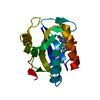
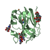
Entry Database : PDB / ID : 5cuhTitle Crystal structure MMP-9 complexes with a constrained hydroxamate based inhibitor LT4 Matrix metalloproteinase-9,Matrix metalloproteinase-9 Keywords / Function / homology Function Domain/homology Component
/ / / / / / / / / / / / / / / / / / / / / / / / / / / / / / / / / / / / / / / / / / / / / / / / / / / / / / / / / / / / / / / / / / / / / / / / / / / / / / / / / / / / / / / / / / / / / / / / / / / / Biological species Homo sapiens (human)Method / / / Resolution : 1.83 Å Authors Tepshi, L. / Vera, L. / Nuti, E. / Rosalia, L. / Rossello, A. / Stura, E.A. Journal : Eur.J.Med.Chem. / Year : 2016Title : Discovery of a new selective inhibitor of A Disintegrin And Metalloprotease 10 (ADAM-10) able to reduce the shedding of NKG2D ligands in Hodgkin's lymphoma cell models.Authors : Camodeca, C. / Nuti, E. / Tepshi, L. / Boero, S. / Tuccinardi, T. / Stura, E.A. / Poggi, A. / Zocchi, M.R. / Rossello, A. History Deposition Jul 24, 2015 Deposition site / Processing site Revision 1.0 Feb 10, 2016 Provider / Type Revision 1.1 Apr 20, 2016 Group Revision 1.2 Sep 27, 2017 Group / Database references / Derived calculationsCategory / diffrn_detector / pdbx_struct_oper_listItem / _diffrn_detector.detector / _pdbx_struct_oper_list.symmetry_operationRevision 1.3 Nov 8, 2023 Group Data collection / Database references ... Data collection / Database references / Derived calculations / Refinement description Category chem_comp_atom / chem_comp_bond ... chem_comp_atom / chem_comp_bond / database_2 / pdbx_initial_refinement_model / pdbx_struct_conn_angle / struct_conn Item _database_2.pdbx_DOI / _database_2.pdbx_database_accession ... _database_2.pdbx_DOI / _database_2.pdbx_database_accession / _pdbx_struct_conn_angle.ptnr1_auth_seq_id / _pdbx_struct_conn_angle.ptnr3_auth_seq_id / _pdbx_struct_conn_angle.value / _struct_conn.pdbx_dist_value / _struct_conn.pdbx_ptnr1_label_alt_id / _struct_conn.ptnr1_auth_asym_id / _struct_conn.ptnr1_auth_comp_id / _struct_conn.ptnr1_auth_seq_id / _struct_conn.ptnr1_label_asym_id / _struct_conn.ptnr1_label_atom_id / _struct_conn.ptnr1_label_comp_id / _struct_conn.ptnr1_label_seq_id / _struct_conn.ptnr2_auth_asym_id / _struct_conn.ptnr2_auth_comp_id / _struct_conn.ptnr2_auth_seq_id / _struct_conn.ptnr2_label_asym_id / _struct_conn.ptnr2_label_atom_id / _struct_conn.ptnr2_label_comp_id
Show all Show less
 Movie
Movie Controller

 Yorodumi
 Open data
 Basic information
 Components
 Keywords
 HYDROLASE / MMP-9 hydroxamate-based inhibitor gelatinase
 Function and homology information
 Authors
 Citation
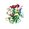
 Structure visualization
 Downloads & links
 Other downloads
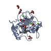

 PDBj
PDBj


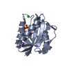
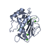
 Assembly

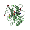


 Mass: 65.409 Da / Num. of mol.: 4 / Source method: obtained synthetically / Formula: Zn
 Mass: 40.078 Da / Num. of mol.: 6 / Source method: obtained synthetically / Formula: Ca
 Mass: 411.499 Da / Num. of mol.: 2 / Source method: obtained synthetically / Formula: C16H21N5O4S2
 Mass: 62.068 Da / Num. of mol.: 4 / Source method: obtained synthetically / Formula: C2H6O2
 Mass: 76.094 Da / Num. of mol.: 2 / Source method: obtained synthetically / Formula: C3H8O2
 Mass: 78.133 Da / Num. of mol.: 1 / Source method: obtained synthetically / Formula: C2H6OS / Comment: DMSO, precipitant*YM
 Sample preparation
 / Beamline: PROXIMA 2 / Wavelength: 0.9801 Å
 Processing