 Movie
Movie Controller

[English] 日本語
 Yorodumi
- PDB-3i8d: The Pairing Geometry of the Hydrophobic Thymine Analog 2,4-Difluo... -
+ Open data

- Basic information
| Entry | Database: PDB / ID: 3i8d | ||||||
|---|---|---|---|---|---|---|---|
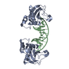
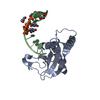
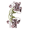
| Title | The Pairing Geometry of the Hydrophobic Thymine Analog 2,4-Difluorotoluene in Duplex DNA as Analyzed by X-ray Crystallography | ||||||
 Components Components |
| ||||||
 Keywords Keywords | HYDROLASE/DNA / RNase-H / RNase-H DNA-complex / 2 / 4-Difluorotoluene / thymine isostere / hydrophobic base /  Endonuclease / Hydrolase / Magnesium / Manganese / Metal-binding / Nuclease / HYDROLASE-DNA COMPLEX Endonuclease / Hydrolase / Magnesium / Manganese / Metal-binding / Nuclease / HYDROLASE-DNA COMPLEX | ||||||
| Function / homology |  Function and homology informationribonuclease H / RNA-DNA hybrid ribonuclease activity / nucleic acid binding / metal ion binding / cytoplasm Function and homology informationribonuclease H / RNA-DNA hybrid ribonuclease activity / nucleic acid binding / metal ion binding / cytoplasmSimilarity search - Function | ||||||
| Biological species |  Bacillus halodurans (bacteria) Bacillus halodurans (bacteria) | ||||||
| Method | X-RAY DIFFRACTION / SYNCHROTRON / using MOLREP / Resolution: 1.61 Å | ||||||
 Authors Authors | Egli, M. / Pallan, P.S. | ||||||
 Citation Citation | Journal: J.Am.Chem.Soc. / Year: 2009 Title: Pairing geometry of the hydrophobic thymine analogue 2,4-difluorotoluene in duplex DNA as analyzed by X-ray crystallography. Authors: Pallan, P.S. / Egli, M. | ||||||
| History |
|
- Structure visualization
| Structure viewer | Molecule: MolmilJmol/JSmol |
|---|
- Downloads & links
-Download
| PDBx/mmCIF format | 3i8d.cif.gz | 90.8 KB | Display | PDBx/mmCIF format |
|---|---|---|---|---|
| PDB format | pdb3i8d.ent.gz | 64.9 KB | Display | PDB format |
| PDBx/mmJSON format | 3i8d.json.gz | Tree view | PDBx/mmJSON format | |
| Others |  Other downloads Other downloads |
-Validation report
| Arichive directory | https://data.pdbj.org/pub/pdb/validation_reports/i8/3i8dftp://data.pdbj.org/pub/pdb/validation_reports/i8/3i8d | HTTPS FTP |
|---|
-Related structure data
| Related structure data |  3d0pS S: Starting model for refinement |
|---|---|
| Similar structure data |


-Links
 PDBj
PDBj


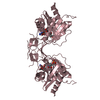
- Assembly
| Deposited unit | 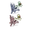
| ||||||||
|---|---|---|---|---|---|---|---|---|---|
| 1 | 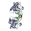
| ||||||||
| 2 | 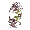
| ||||||||
| Unit cell |
| ||||||||
| Details | The second part of the biological assembly is generated by the two fold axis rotation of chain A and B by: -x, y, -z. |
-Components
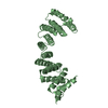
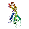
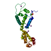
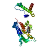
| #1: Protein | / RNase H Mass: 15243.206 Da / Num. of mol.: 2 / Fragment: RNase-H / Mutation: D132N Source method: isolated from a genetically manipulated source Source: (gene. exp.) Bacillus halodurans (bacteria)Gene: BACILLUS HALODURANS, BH0863, RNase-H D132N mutant, rnhA Production host: Escherichia coli (E. coli) / References: UniProt: Q9KEI9, ribonuclease H#2: DNA chain | Mass: 3665.398 Da / Num. of mol.: 2 / Source method: obtained synthetically #3: Chemical | ChemComp-GOL / Glycerol  Mass: 92.094 Da / Num. of mol.: 4 / Source method: obtained synthetically / Formula: C3H8O3 Mass: 92.094 Da / Num. of mol.: 4 / Source method: obtained synthetically / Formula: C3H8O3#4: Water | ChemComp-HOH / | Water Mass: 18.015 Da / Num. of mol.: 231 / Source method: isolated from a natural source / Formula: H2O Mass: 18.015 Da / Num. of mol.: 231 / Source method: isolated from a natural source / Formula: H2O |
|---|
-Experimental details
-Experiment
| Experiment | Method: X-RAY DIFFRACTION / Number of used crystals: 1 |
|---|
- Sample preparation
| Crystal | Density Matthews: 2.82 Å3/Da / Density % sol: 56.37 % |
|---|---|
| Crystal grow | Temperature: 291 K / Method: vapor diffusion, sitting drop / pH: 4.6 Details: 0.1 M NaOAc 3H2O (pH 4.6) and 8% (w/v) PEG 4000 , VAPOR DIFFUSION, SITTING DROP, temperature 291K |
-Data collection
| Diffraction | Mean temperature: 100 K |
|---|---|
| Diffraction source | Source: SYNCHROTRON / Site: APS  / Beamline: 21-ID-F / Wavelength: 0.9787 Å / Beamline: 21-ID-F / Wavelength: 0.9787 Å |
| Detector | Type: MARMOSAIC 225 mm CCD / Detector: CCD / Date: Nov 17, 2008 |
| Radiation | Protocol: SINGLE WAVELENGTH / Monochromatic (M) / Laue (L): M / Scattering type: x-ray |
| Radiation wavelength | Wavelength: 0.9787 Å / Relative weight: 1 |
| Reflection | Resolution: 1.61→50 Å / Num. all: 53844 / Num. obs: 52821 / % possible obs: 98.1 % / Observed criterion σ(F): 0 / Observed criterion σ(I): 0 / Redundancy: 7.5 % / Rmerge(I) obs: 0.053 / Net I/σ(I): 39.4 |
| Reflection shell | Resolution: 1.61→1.64 Å / Redundancy: 4.6 % / Rmerge(I) obs: 0.56 / Mean I/σ(I) obs: 1.7 / Num. measured all: 2686 / Num. unique all: 2203 |
- Processing
| Software |
| |||||||||||||||||||||||||||||||||||||||||||||||||||||||||||||||||||||||||||||||||||||||||||||||||||||||||||||||||||||||||||||
|---|---|---|---|---|---|---|---|---|---|---|---|---|---|---|---|---|---|---|---|---|---|---|---|---|---|---|---|---|---|---|---|---|---|---|---|---|---|---|---|---|---|---|---|---|---|---|---|---|---|---|---|---|---|---|---|---|---|---|---|---|---|---|---|---|---|---|---|---|---|---|---|---|---|---|---|---|---|---|---|---|---|---|---|---|---|---|---|---|---|---|---|---|---|---|---|---|---|---|---|---|---|---|---|---|---|---|---|---|---|---|---|---|---|---|---|---|---|---|---|---|---|---|---|---|---|---|
| Refinement | Method to determine structure: using MOLREP Starting model: PDB ID 3D0P Resolution: 1.61→49.39 Å / Cor.coef. Fo:Fc: 0.966 / Cor.coef. Fo:Fc free: 0.954 / SU B: 4.099 / SU ML: 0.064 / TLS residual ADP flag: LIKELY RESIDUAL / Cross valid method: THROUGHOUT / ESU R: 0.088 / ESU R Free: 0.094 / Stereochemistry target values: MAXIMUM LIKELIHOOD / Details: TLS refinement in REFMAC was used
| |||||||||||||||||||||||||||||||||||||||||||||||||||||||||||||||||||||||||||||||||||||||||||||||||||||||||||||||||||||||||||||
| Solvent computation | Ion probe radii: 0.8 Å / Shrinkage radii: 0.8 Å / VDW probe radii: 1.4 Å / Solvent model: MASK | |||||||||||||||||||||||||||||||||||||||||||||||||||||||||||||||||||||||||||||||||||||||||||||||||||||||||||||||||||||||||||||
| Displacement parameters | Biso mean: 16.68 Å2
| |||||||||||||||||||||||||||||||||||||||||||||||||||||||||||||||||||||||||||||||||||||||||||||||||||||||||||||||||||||||||||||
| Refinement step | Cycle: LAST / Resolution: 1.61→49.39 Å
| |||||||||||||||||||||||||||||||||||||||||||||||||||||||||||||||||||||||||||||||||||||||||||||||||||||||||||||||||||||||||||||
| Refine LS restraints |
| |||||||||||||||||||||||||||||||||||||||||||||||||||||||||||||||||||||||||||||||||||||||||||||||||||||||||||||||||||||||||||||
| LS refinement shell | Resolution: 1.613→1.655 Å / Total num. of bins used: 20
| |||||||||||||||||||||||||||||||||||||||||||||||||||||||||||||||||||||||||||||||||||||||||||||||||||||||||||||||||||||||||||||
| Refinement TLS params. | Method: refined / Refine-ID: X-RAY DIFFRACTION
| |||||||||||||||||||||||||||||||||||||||||||||||||||||||||||||||||||||||||||||||||||||||||||||||||||||||||||||||||||||||||||||
| Refinement TLS group |
|
