 Movie
Movie Controller


+ Open data

- Basic information
| Entry | Database: PDB / ID: 1xtq | ||||||
|---|---|---|---|---|---|---|---|
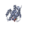
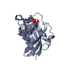
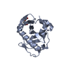
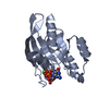
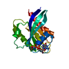
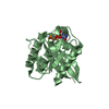
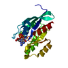
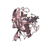
| Title | Structure of small GTPase human Rheb in complex with GDP | ||||||
 Components Components | GTP-binding protein Rheb | ||||||
 Keywords Keywords |  SIGNALING PROTEIN / beta saddle / P-loop SIGNALING PROTEIN / beta saddle / P-loop | ||||||
| Function / homology |  Function and homology information Function and homology informationregulation of type B pancreatic cell development / MTOR signalling / Amino acids regulate mTORC1 / Energy dependent regulation of mTOR by LKB1-AMPK / negative regulation of cold-induced thermogenesis / Macroautophagy / small GTPase-mediated signal transduction / positive regulation of oligodendrocyte differentiation / protein kinase activator activity / oligodendrocyte differentiation ...regulation of type B pancreatic cell development / MTOR signalling / Amino acids regulate mTORC1 / Energy dependent regulation of mTOR by LKB1-AMPK / negative regulation of cold-induced thermogenesis / Macroautophagy / small GTPase-mediated signal transduction / positive regulation of oligodendrocyte differentiation / protein kinase activator activity / oligodendrocyte differentiation / mTORC1-mediated signalling / cellular response to nutrient levels / positive regulation of TOR signaling / regulation of macroautophagy / endomembrane system / positive regulation of TORC1 signaling / protein serine/threonine kinase activator activity / Regulation of PTEN gene transcription / TP53 Regulates Metabolic Genes / Hydrolases; Acting on acid anhydrides; Acting on GTP to facilitate cellular and subcellular movement / spliceosomal complex / GDP binding / postsynaptic density / regulation of cell cycle / lysosomal membrane / Golgi membrane / GTPase activity / endoplasmic reticulum membrane / GTP binding / protein kinase binding / magnesium ion binding / signal transduction / extracellular exosome / membrane / plasma membrane / cytosolSimilarity search - Function | ||||||
| Biological species |  Homo sapiens (human) Homo sapiens (human) | ||||||
| Method | X-RAY DIFFRACTION / MOLECULAR REPLACEMENT / Resolution: 2 Å | ||||||
 Authors Authors | Yu, Y. / Ding, J. | ||||||
 Citation Citation | Journal: J.Biol.Chem. / Year: 2005 Title: Structural Basis for the Unique Biological Function of Small GTPase RHEB Authors: Yu, Y. / Li, S. / Xu, X. / Li, Y. / Guan, K. / Arnold, E. / Ding, J. | ||||||
| History |
|
- Structure visualization
| Structure viewer | Molecule: MolmilJmol/JSmol |
|---|
- Downloads & links
-Download
| PDBx/mmCIF format | 1xtq.cif.gz | 53.1 KB | Display | PDBx/mmCIF format |
|---|---|---|---|---|
| PDB format | pdb1xtq.ent.gz | 36.2 KB | Display | PDB format |
| PDBx/mmJSON format | 1xtq.json.gz | Tree view | PDBx/mmJSON format | |
| Others |  Other downloads Other downloads |
-Validation report
| Arichive directory | https://data.pdbj.org/pub/pdb/validation_reports/xt/1xtqftp://data.pdbj.org/pub/pdb/validation_reports/xt/1xtq | HTTPS FTP |
|---|
-Related structure data
| Related structure data | 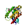 1xtrC 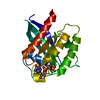 1xtsC  1guaS 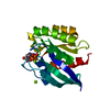 1kaoS C: citing same article ( S: Starting model for refinement |
|---|---|
| Similar structure data |


-Links
 PDBj
PDBj


- Assembly
| Deposited unit | 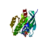
| ||||||||
|---|---|---|---|---|---|---|---|---|---|
| 1 |
| ||||||||
| Unit cell |
|
-Components
| #1: Protein | Mass: 20148.965 Da / Num. of mol.: 1 / Fragment: GTPase domain Source method: isolated from a genetically manipulated source Source: (gene. exp.) Homo sapiens (human) / Gene: Rheb / Plasmid: pET22b(+) / Species (production host): Escherichia coli / Production host:  Escherichia coli BL21(DE3) (bacteria) / Strain (production host): BL21(DE3) / References: UniProt: Q15382 Escherichia coli BL21(DE3) (bacteria) / Strain (production host): BL21(DE3) / References: UniProt: Q15382 |
|---|---|
| #2: Chemical | ChemComp-MG /   Mass: 24.305 Da / Num. of mol.: 1 / Source method: obtained synthetically / Formula: Mg Mass: 24.305 Da / Num. of mol.: 1 / Source method: obtained synthetically / Formula: Mg |
| #3: Chemical | ChemComp-GDP / Guanosine diphosphate  Type: RNA linking / Mass: 443.201 Da / Num. of mol.: 1 / Source method: obtained synthetically / Formula: C10H15N5O11P2 / Comment: GDP, energy-carrying molecule*YM Type: RNA linking / Mass: 443.201 Da / Num. of mol.: 1 / Source method: obtained synthetically / Formula: C10H15N5O11P2 / Comment: GDP, energy-carrying molecule*YM |
| #4: Water | ChemComp-HOH / Water Mass: 18.015 Da / Num. of mol.: 164 / Source method: isolated from a natural source / Formula: H2O Mass: 18.015 Da / Num. of mol.: 164 / Source method: isolated from a natural source / Formula: H2O |
-Experimental details
-Experiment
| Experiment | Method: X-RAY DIFFRACTION / Number of used crystals: 1 |
|---|
- Sample preparation
| Crystal | Density Matthews: 2.1 Å3/Da / Density % sol: 40.1 % |
|---|---|
| Crystal grow | Temperature: 277 K / Method: vapor diffusion, hanging drop / pH: 4.6 Details: potassium dihydrogenphosphate, PEG 8000, pH 4.6, VAPOR DIFFUSION, HANGING DROP, temperature 277K |
-Data collection
| Diffraction | Mean temperature: 100 K |
|---|---|
| Diffraction source | Source: ROTATING ANODE / Type: RIGAKU / Wavelength: 1.5418 Å |
| Detector | Type: RIGAKU RAXIS IV / Detector: IMAGE PLATE / Date: Jan 30, 2004 / Details: osmic mirrors |
| Radiation | Monochromator: Ni filter / Protocol: SINGLE WAVELENGTH / Monochromatic (M) / Laue (L): M / Scattering type: x-ray |
| Radiation wavelength | Wavelength: 1.5418 Å / Relative weight: 1 |
| Reflection | Resolution: 2→14.8 Å / Num. all: 11457 / Num. obs: 11457 / % possible obs: 98.9 % / Observed criterion σ(F): 0 / Observed criterion σ(I): 0 / Redundancy: 2.6 % / Biso Wilson estimate: 20.1 Å2 / Rmerge(I) obs: 0.029 / Net I/σ(I): 18.6 |
| Reflection shell | Resolution: 2→2.07 Å / Redundancy: 2.5 % / Rmerge(I) obs: 0.122 / Mean I/σ(I) obs: 4.9 / Num. unique all: 1126 / % possible all: 98.7 |
- Processing
| Software |
| |||||||||||||||||||||||||||||||||||||||||||||||||
|---|---|---|---|---|---|---|---|---|---|---|---|---|---|---|---|---|---|---|---|---|---|---|---|---|---|---|---|---|---|---|---|---|---|---|---|---|---|---|---|---|---|---|---|---|---|---|---|---|---|---|
| Refinement | Method to determine structure: MOLECULAR REPLACEMENT Starting model: PDB entries 1KAO, 1GUA Resolution: 2→14.8 Å / Isotropic thermal model: isotropic / Cross valid method: THROUGHOUT / σ(F): 0 / σ(I): 0 / Stereochemistry target values: Engh & Huber / Details: Used weighted full matrix least squares procedure.
| |||||||||||||||||||||||||||||||||||||||||||||||||
| Displacement parameters | Biso mean: 30.2 Å2 | |||||||||||||||||||||||||||||||||||||||||||||||||
| Refine analyze |
| |||||||||||||||||||||||||||||||||||||||||||||||||
| Refinement step | Cycle: LAST / Resolution: 2→14.8 Å
| |||||||||||||||||||||||||||||||||||||||||||||||||
| Refine LS restraints |
| |||||||||||||||||||||||||||||||||||||||||||||||||
| LS refinement shell |
|
