 Movie
Movie Controller


+ Open data

- Basic information
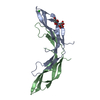
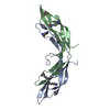
| Entry | Database: PDB / ID: 3lqm | ||||||
|---|---|---|---|---|---|---|---|
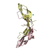
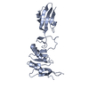
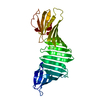
| Title | Structure of the IL-10R2 Common Chain | ||||||
 Components Components | Interleukin-10 receptor subunit beta | ||||||
 Keywords Keywords | PROTEIN BINDING / IL-10R2 / receptor / common chain / cytokine / IL-10 / IL-22 / IL-26 / IL-28 / IL-29 / Disulfide bond / Glycoprotein / Membrane / Phosphoprotein / Polymorphism / Transmembrane | ||||||
| Function / homology |  Function and homology informationinterleukin-10 receptor activity / interleukin-28 receptor complex / positive regulation of cellular respiration / type III interferon-mediated signaling pathway / Other interleukin signaling / Interleukin-20 family signaling / Interleukin-10 signaling / positive regulation of receptor signaling pathway via JAK-STAT / cellular response to virus / cytokine-mediated signaling pathway ...interleukin-10 receptor activity / interleukin-28 receptor complex / positive regulation of cellular respiration / type III interferon-mediated signaling pathway / Other interleukin signaling / Interleukin-20 family signaling / Interleukin-10 signaling / positive regulation of receptor signaling pathway via JAK-STAT / cellular response to virus / cytokine-mediated signaling pathway / signaling receptor activity / defense response to virus / immune response / inflammatory response / signal transduction / membrane / plasma membrane Function and homology informationinterleukin-10 receptor activity / interleukin-28 receptor complex / positive regulation of cellular respiration / type III interferon-mediated signaling pathway / Other interleukin signaling / Interleukin-20 family signaling / Interleukin-10 signaling / positive regulation of receptor signaling pathway via JAK-STAT / cellular response to virus / cytokine-mediated signaling pathway ...interleukin-10 receptor activity / interleukin-28 receptor complex / positive regulation of cellular respiration / type III interferon-mediated signaling pathway / Other interleukin signaling / Interleukin-20 family signaling / Interleukin-10 signaling / positive regulation of receptor signaling pathway via JAK-STAT / cellular response to virus / cytokine-mediated signaling pathway / signaling receptor activity / defense response to virus / immune response / inflammatory response / signal transduction / membrane / plasma membraneSimilarity search - Function | ||||||
| Biological species |  Homo sapiens (human) Homo sapiens (human) | ||||||
| Method | X-RAY DIFFRACTION / SYNCHROTRON / MAD / Resolution: 2.14 Å | ||||||
 Authors Authors | Yoon, S.I. / Walter, M.R. | ||||||
 Citation Citation | Journal: Structure / Year: 2010 Title: Structure and mechanism of receptor sharing by the IL-10R2 common chain. Authors: Yoon, S.I. / Jones, B.C. / Logsdon, N.J. / Harris, B.D. / Deshpande, A. / Radaeva, S. / Halloran, B.A. / Gao, B. / Walter, M.R. | ||||||
| History |
|
- Structure visualization
| Structure viewer | Molecule: MolmilJmol/JSmol |
|---|
- Downloads & links
-Download
| PDBx/mmCIF format | 3lqm.cif.gz | 94.9 KB | Display | PDBx/mmCIF format |
|---|---|---|---|---|
| PDB format | pdb3lqm.ent.gz | 77.4 KB | Display | PDB format |
| PDBx/mmJSON format | 3lqm.json.gz | Tree view | PDBx/mmJSON format | |
| Others |  Other downloads Other downloads |
-Validation report
| Arichive directory | https://data.pdbj.org/pub/pdb/validation_reports/lq/3lqmftp://data.pdbj.org/pub/pdb/validation_reports/lq/3lqm | HTTPS FTP |
|---|
-Related structure data
| Similar structure data |
|---|


-Links
 PDBj
PDBj


- Assembly
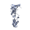
| Deposited unit | 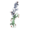
| ||||||||
|---|---|---|---|---|---|---|---|---|---|
| 1 | 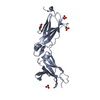
| ||||||||
| 2 | 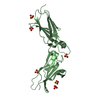
| ||||||||
| Unit cell |
|
-Components
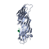
| #1: Protein | / IL-10 receptor subunit beta / IL-10R subunit beta / IL-10RB / Interleukin-10 receptor subunit 2 / ...IL-10 receptor subunit beta / IL-10R subunit beta / IL-10RB / Interleukin-10 receptor subunit 2 / IL-10R subunit 2 / IL-10R2 / Cytokine receptor family 2 member 4 / CRF2-4 / Cytokine receptor class-II member 4 Mass: 23583.361 Da / Num. of mol.: 2 / Fragment: Extracellular Domain / Mutation: N49Q, N68Q, N102Q, C106S, S126C, N161Q Source method: isolated from a genetically manipulated source Source: (gene. exp.) Homo sapiens (human) / Production host: Spodoptera frugiperda / References: UniProt: Q08334#2: Chemical | ChemComp-SO4 / Sulfate  Mass: 96.063 Da / Num. of mol.: 10 / Source method: obtained synthetically / Formula: SO4 Mass: 96.063 Da / Num. of mol.: 10 / Source method: obtained synthetically / Formula: SO4#3: Chemical | ChemComp-GOL / | Glycerol  Mass: 92.094 Da / Num. of mol.: 1 / Source method: obtained synthetically / Formula: C3H8O3 Mass: 92.094 Da / Num. of mol.: 1 / Source method: obtained synthetically / Formula: C3H8O3#4: Water | ChemComp-HOH / | Water Mass: 18.015 Da / Num. of mol.: 212 / Source method: isolated from a natural source / Formula: H2O Mass: 18.015 Da / Num. of mol.: 212 / Source method: isolated from a natural source / Formula: H2O |
|---|
-Experimental details
-Experiment
| Experiment | Method: X-RAY DIFFRACTION / Number of used crystals: 1 |
|---|
- Sample preparation
| Crystal | Density Matthews: 4.02 Å3/Da / Density % sol: 69.38 % |
|---|---|
| Crystal grow | Temperature: 298 K / Method: vapor diffusion, hanging drop / pH: 7.5 Details: pH 7.5, VAPOR DIFFUSION, HANGING DROP, temperature 298K |
-Data collection
| Diffraction | Mean temperature: 100 K |
|---|---|
| Diffraction source | Source: SYNCHROTRON / Site: APS  / Beamline: 22-ID / Wavelength: 1 Å / Beamline: 22-ID / Wavelength: 1 Å |
| Detector | Type: ADSC QUANTUM 315 / Detector: CCD / Date: Oct 6, 2006 |
| Radiation | Protocol: MAD / Monochromatic (M) / Laue (L): M / Scattering type: x-ray |
| Radiation wavelength | Wavelength: 1 Å / Relative weight: 1 |
| Reflection | Resolution: 2.14→50 Å / Num. all: 41552 / Num. obs: 40491 / % possible obs: 97.5 % / Observed criterion σ(F): 0 / Observed criterion σ(I): -3 / Redundancy: 11 % / Rsym value: 0.071 / Net I/σ(I): 50 |
- Processing
| Software |
| |||||||||||||||||||||
|---|---|---|---|---|---|---|---|---|---|---|---|---|---|---|---|---|---|---|---|---|---|---|
| Refinement | Method to determine structure: MAD / Resolution: 2.14→50 Å / Cross valid method: THROUGHOUT / σ(F): 0 / σ(I): -3 / Stereochemistry target values: Engh & Huber
| |||||||||||||||||||||
| Displacement parameters | Biso mean: 37.3 Å2 | |||||||||||||||||||||
| Refinement step | Cycle: LAST / Resolution: 2.14→50 Å
|
