| Entry | Database: PDB / ID: 2vh1
|
|---|
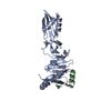
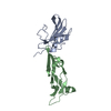
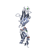
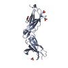
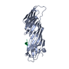
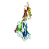
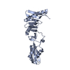
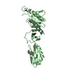
| Title | Crystal structure of bacterial cell division protein FtsQ from E.coli |
|---|
 Components Components | CELL DIVISION PROTEIN FTSQ |
|---|
 Keywords Keywords | CELL CYCLE / FTSQ / POTRA / MEMBRANE / SEPTATION / CELL DIVISION / TRANSMEMBRANE / INNER MEMBRANE |
|---|
| Function / homology |  Function and homology information Function and homology information
FtsQBL complex / divisome complex / FtsZ-dependent cytokinesis / division septum assembly / cell division site / cell division / identical protein binding / plasma membraneSimilarity search - Function Cell division protein FtsQ/DivIB / Cell division protein FtsQ, C-terminal / Cell division protein FtsQ / Cell division protein FtsQ/DivIB, C-terminal / POTRA domain, FtsQ-type / Cell division protein FtsQ/DivIB, C-terminal / POTRA domain, FtsQ-type / membrane protein fhac / POTRA domain / POTRA domain profile. ...Cell division protein FtsQ/DivIB / Cell division protein FtsQ, C-terminal / Cell division protein FtsQ / Cell division protein FtsQ/DivIB, C-terminal / POTRA domain, FtsQ-type / Cell division protein FtsQ/DivIB, C-terminal / POTRA domain, FtsQ-type / membrane protein fhac / POTRA domain / POTRA domain profile. / Ubiquitin-like (UB roll) / Roll / Rossmann fold / 3-Layer(aba) Sandwich / Alpha BetaSimilarity search - Domain/homology |
|---|
| Biological species |   ESCHERICHIA COLI (E. coli) ESCHERICHIA COLI (E. coli) |
|---|
| Method | X-RAY DIFFRACTION / SYNCHROTRON / MAD / Resolution: 2.7 Å |
|---|
 Authors Authors | van den Ent, F. / Vinkenvleugel, T. / Ind, A. / West, P. / Veprintsev, D. / Naninga, N. / den Blaauwen, T. / Lowe, J. |
|---|
 Citation Citation | Journal: Mol.Microbiol. / Year: 2008
Title: Structural and Mutational Analysis of Cell Division Protein Ftsq
Authors: van den Ent, F. / Vinkenvleugel, T. / Ind, A. / West, P. / Veprintsev, D. / Naninga, N. / den Blaauwen, T. / Lowe, J. |
|---|
| History | | Deposition | Nov 16, 2007 | Deposition site: PDBE / Processing site: PDBE |
|---|
| Revision 1.0 | Mar 11, 2008 | Provider: repository / Type: Initial release |
|---|
| Revision 1.1 | Jul 13, 2011 | Group: Advisory / Version format compliance |
|---|
| Revision 1.2 | Jan 24, 2018 | Group: Database references / Source and taxonomy / Category: citation_author / entity_src_gen
Item: _citation_author.name / _entity_src_gen.pdbx_host_org_ncbi_taxonomy_id ..._citation_author.name / _entity_src_gen.pdbx_host_org_ncbi_taxonomy_id / _entity_src_gen.pdbx_host_org_scientific_name / _entity_src_gen.pdbx_host_org_strain / _entity_src_gen.pdbx_host_org_variant |
|---|
| Revision 1.3 | May 8, 2024 | Group: Data collection / Database references ...Data collection / Database references / Other / Refinement description
Category: chem_comp_atom / chem_comp_bond ...chem_comp_atom / chem_comp_bond / database_2 / pdbx_database_status / struct_ncs_dom_lim
Item: _database_2.pdbx_DOI / _database_2.pdbx_database_accession ..._database_2.pdbx_DOI / _database_2.pdbx_database_accession / _pdbx_database_status.status_code_sf / _struct_ncs_dom_lim.beg_auth_comp_id / _struct_ncs_dom_lim.beg_label_asym_id / _struct_ncs_dom_lim.beg_label_comp_id / _struct_ncs_dom_lim.beg_label_seq_id / _struct_ncs_dom_lim.end_auth_comp_id / _struct_ncs_dom_lim.end_label_asym_id / _struct_ncs_dom_lim.end_label_comp_id / _struct_ncs_dom_lim.end_label_seq_id |
|---|
|
|---|
 Movie
Movie Controller

 Yorodumi
 Open data
 Basic information
 Structure visualization
 Downloads & links
 Other downloads
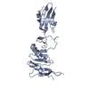

 PDBj
PDBj Assembly
 Sample preparation
 / Beamline: ID23-1 / Wavelength: 0.9392
  Processing