 Movie
Movie Controller

[English] 日本語
 Yorodumi
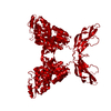
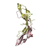
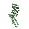
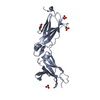
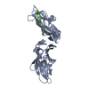
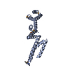
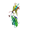
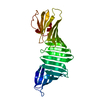
- PDB-5bq8: Crystal structure of Norrin, a Wnt signalling activator, Crystal ... -
+ Open data

- Basic information
| Entry | Database: PDB / ID: 5bq8 | ||||||||||||
|---|---|---|---|---|---|---|---|---|---|---|---|---|---|
| Title | Crystal structure of Norrin, a Wnt signalling activator, Crystal Form II | ||||||||||||
 Components Components | (Norrin ) x 3 ) x 3 | ||||||||||||
 Keywords Keywords | SIGNALING PROTEIN / Wnt signalling pathway / Norrie disease protein / cystine-knot like growth factor / ligand for Frizzled 4 receptor | ||||||||||||
| Function / homology |  Function and homology information Function and homology informationretina blood vessel maintenance / cone retinal bipolar cell differentiation / Norrin signaling pathway / extracellular matrix-cell signaling / re-entry into mitotic cell cycle / retinal blood vessel morphogenesis / retinal rod cell differentiation / ubiquitin-dependent endocytosis / retina layer formation / retinal pigment epithelium development ...retina blood vessel maintenance / cone retinal bipolar cell differentiation / Norrin signaling pathway / extracellular matrix-cell signaling / re-entry into mitotic cell cycle / retinal blood vessel morphogenesis / retinal rod cell differentiation / ubiquitin-dependent endocytosis / retina layer formation / retinal pigment epithelium development / establishment of blood-retinal barrier / microglial cell proliferation / glycine metabolic process / endothelial cell differentiation / dendritic spine development / establishment of blood-brain barrier / protein targeting to lysosome / vacuole organization / microglia differentiation / frizzled binding / optic nerve development / retinal ganglion cell axon guidance / lens development in camera-type eye / exploration behavior / action potential / smoothened signaling pathway / decidualization / canonical Wnt signaling pathway / response to axon injury / blood vessel remodeling / glutathione metabolic process / tricarboxylic acid cycle / visual perception / transforming growth factor beta receptor signaling pathway / phosphatidylinositol 3-kinase/protein kinase B signal transduction / cytokine activity / mitotic cell cycle / nervous system development / cellular response to hypoxia / angiogenesis / neuron apoptotic process / collagen-containing extracellular matrix / transcription by RNA polymerase II / protein ubiquitination / inflammatory response / positive regulation of DNA-templated transcription / cell surface / protein homodimerization activity / extracellular spaceSimilarity search - Function | ||||||||||||
| Biological species |  Homo sapiens (human) Homo sapiens (human) | ||||||||||||
| Method | X-RAY DIFFRACTION / SYNCHROTRON / MOLECULAR REPLACEMENT / Resolution: 2 Å | ||||||||||||
 Authors Authors | Chang, T.-H. / Hsieh, F.-L. / Harlos, K. / Jones, E.Y. | ||||||||||||
| Funding support |  United Kingdom, 3items United Kingdom, 3items
| ||||||||||||
 Citation Citation | Journal: Elife / Year: 2015 Title: Structure and functional properties of Norrin mimic Wnt for signalling with Frizzled4, Lrp5/6, and proteoglycan. Authors: Chang, T.H. / Hsieh, F.L. / Zebisch, M. / Harlos, K. / Elegheert, J. / Jones, E.Y. | ||||||||||||
| History |
|
- Structure visualization
| Structure viewer | Molecule: MolmilJmol/JSmol |
|---|
- Downloads & links
-Download
| PDBx/mmCIF format | 5bq8.cif.gz | 98.7 KB | Display | PDBx/mmCIF format |
|---|---|---|---|---|
| PDB format | pdb5bq8.ent.gz | 75.1 KB | Display | PDB format |
| PDBx/mmJSON format | 5bq8.json.gz | Tree view | PDBx/mmJSON format | |
| Others |  Other downloads Other downloads |
-Validation report
| Arichive directory | https://data.pdbj.org/pub/pdb/validation_reports/bq/5bq8ftp://data.pdbj.org/pub/pdb/validation_reports/bq/5bq8 | HTTPS FTP |
|---|
-Related structure data
| Related structure data | 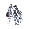 5bpbC 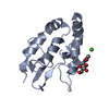 5bpqC 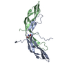 5bpuSC 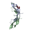 5bqbC 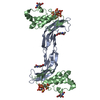 5bqcC 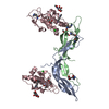 5bqeC S: Starting model for refinement C: citing same article ( |
|---|---|
| Similar structure data |


-Links
 PDBj
PDBj

- Assembly
| Deposited unit | 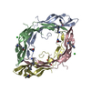
| ||||||||
|---|---|---|---|---|---|---|---|---|---|
| 1 | 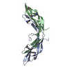
| ||||||||
| 2 | 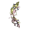
| ||||||||
| Unit cell |
|
-Components
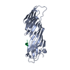
-Protein , 3 types, 4 molecules ACBD
| #1: Protein | / Norrie disease protein / X-linked exudative vitreoretinopathy 2 protein Mass: 13668.798 Da / Num. of mol.: 2 / Fragment: UNP residues 25-133 Source method: isolated from a genetically manipulated source Source: (gene. exp.) Homo sapiens (human) / Gene: NDP, EVR2 / Plasmid: Plasmid / Details (production host): pHLIgK-STR-8H-SUMO-1D4 / Cell line (production host): HEK293T / Production host: Homo sapiens (human) / References: UniProt: Q00604#2: Protein | | / Norrie disease protein / X-linked exudative vitreoretinopathy 2 proteinMass: 13695.845 Da / Num. of mol.: 1 / Fragment: UNP residues 25-133 Source method: isolated from a genetically manipulated source Source: (gene. exp.) Homo sapiens (human) / Gene: NDP, EVR2 / Plasmid: Plasmid / Details (production host): pHLIgK-STR-8H-SUMO-1D4 / Cell line (production host): HEK293T / Production host: Homo sapiens (human) / References: UniProt: Q00604#3: Protein | | / Norrie disease protein / X-linked exudative vitreoretinopathy 2 proteinMass: 13722.891 Da / Num. of mol.: 1 / Fragment: UNP residues 25-133 Source method: isolated from a genetically manipulated source Source: (gene. exp.) Homo sapiens (human) / Gene: NDP, EVR2 / Plasmid: Plasmid / Details (production host): pHLIgK-STR-8H-SUMO-1D4 / Cell line (production host): HEK293T / Production host: Homo sapiens (human) / References: UniProt: Q00604 |
|---|
-Non-polymers , 3 types, 126 molecules 


| #4: Chemical | Chloride Mass: 35.453 Da / Num. of mol.: 3 / Source method: obtained synthetically / Formula: Cl Mass: 35.453 Da / Num. of mol.: 3 / Source method: obtained synthetically / Formula: Cl#5: Chemical | ChemComp-PEG / | Diethylene glycol Mass: 106.120 Da / Num. of mol.: 1 / Source method: obtained synthetically / Formula: C4H10O3 Mass: 106.120 Da / Num. of mol.: 1 / Source method: obtained synthetically / Formula: C4H10O3#6: Water | ChemComp-HOH / | WaterMass: 18.015 Da / Num. of mol.: 122 / Source method: isolated from a natural source / Formula: H2O |
|---|
-Experimental details
-Experiment
| Experiment | Method: X-RAY DIFFRACTION |
|---|
- Sample preparation
| Crystal | Density Matthews: 2.41 Å3/Da / Density % sol: 49.1 % |
|---|---|
| Crystal grow | Temperature: 294 K / Method: vapor diffusion, sitting drop Details: 0.1 sodium acetate, pH 5.0, 5% PGA-LM, 4% PEG2000MME, 24% PEG550MME |
-Data collection
| Diffraction | Mean temperature: 100 K |
|---|---|
| Diffraction source | Source: SYNCHROTRON / Site: Diamond / Beamline: I04 / Wavelength: 0.97949 Å |
| Detector | Type: DECTRIS PILATUS 6M / Detector: PIXEL / Date: Sep 15, 2013 |
| Radiation | Protocol: SINGLE WAVELENGTH / Monochromatic (M) / Laue (L): M / Scattering type: x-ray |
| Radiation wavelength | Wavelength: 0.97949 Å / Relative weight: 1 |
| Reflection | Resolution: 2→33.65 Å / Num. obs: 36272 / % possible obs: 100 % / Redundancy: 5.6 % / Rmerge(I) obs: 0.089 / Net I/σ(I): 9.1 |
| Reflection shell | Resolution: 2→2.05 Å / Redundancy: 5.8 % / Rmerge(I) obs: 1.289 / Mean I/σ(I) obs: 1.7 / % possible all: 100 |
- Processing
| Software |
| |||||||||||||||||||||||||||||||||||||||||||||||||||||||||||||||||||||||||||||||||||||||||||||||||||||||||||||||||||||||||||||
|---|---|---|---|---|---|---|---|---|---|---|---|---|---|---|---|---|---|---|---|---|---|---|---|---|---|---|---|---|---|---|---|---|---|---|---|---|---|---|---|---|---|---|---|---|---|---|---|---|---|---|---|---|---|---|---|---|---|---|---|---|---|---|---|---|---|---|---|---|---|---|---|---|---|---|---|---|---|---|---|---|---|---|---|---|---|---|---|---|---|---|---|---|---|---|---|---|---|---|---|---|---|---|---|---|---|---|---|---|---|---|---|---|---|---|---|---|---|---|---|---|---|---|---|---|---|---|
| Refinement | Method to determine structure: MOLECULAR REPLACEMENT Starting model: 5BPU Resolution: 2→33.65 Å / SU ML: 0.29 / Cross valid method: FREE R-VALUE / σ(F): 1.34 / Phase error: 31.27 / Stereochemistry target values: ML
| |||||||||||||||||||||||||||||||||||||||||||||||||||||||||||||||||||||||||||||||||||||||||||||||||||||||||||||||||||||||||||||
| Solvent computation | Shrinkage radii: 0.9 Å / VDW probe radii: 1.11 Å / Solvent model: FLAT BULK SOLVENT MODEL | |||||||||||||||||||||||||||||||||||||||||||||||||||||||||||||||||||||||||||||||||||||||||||||||||||||||||||||||||||||||||||||
| Refinement step | Cycle: LAST / Resolution: 2→33.65 Å
| |||||||||||||||||||||||||||||||||||||||||||||||||||||||||||||||||||||||||||||||||||||||||||||||||||||||||||||||||||||||||||||
| Refine LS restraints |
| |||||||||||||||||||||||||||||||||||||||||||||||||||||||||||||||||||||||||||||||||||||||||||||||||||||||||||||||||||||||||||||
| LS refinement shell |
| |||||||||||||||||||||||||||||||||||||||||||||||||||||||||||||||||||||||||||||||||||||||||||||||||||||||||||||||||||||||||||||
| Refinement TLS params. | Method: refined / Refine-ID: X-RAY DIFFRACTION
| |||||||||||||||||||||||||||||||||||||||||||||||||||||||||||||||||||||||||||||||||||||||||||||||||||||||||||||||||||||||||||||
| Refinement TLS group |
|
