 Movie
Movie Controller

[English] 日本語
 Yorodumi
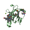
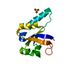
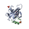
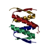
- PDB-5bpq: Crystal structure of the cysteine-rich domain of human Frizzled 4... -
+ Open data

- Basic information
| Entry | Database: PDB / ID: 5bpq | ||||||||||||
|---|---|---|---|---|---|---|---|---|---|---|---|---|---|
| Title | Crystal structure of the cysteine-rich domain of human Frizzled 4 - Crystal Form II | ||||||||||||
 Components Components | Frizzled-4 | ||||||||||||
 Keywords Keywords | SIGNALING PROTEIN / Wnt signalling pathway / Glycoprotein / G protein coupled receptor / receptor for Norrin recognition | ||||||||||||
| Function / homology |  Function and homology information Function and homology informationcerebellum vasculature morphogenesis / Wnt signaling pathway, calcium modulating pathway / Norrin signaling pathway / extracellular matrix-cell signaling / progesterone secretion / retinal blood vessel morphogenesis / locomotion involved in locomotory behavior / retina vasculature morphogenesis in camera-type eye / regulation of vascular endothelial growth factor receptor signaling pathway / Signaling by RNF43 mutants ...cerebellum vasculature morphogenesis / Wnt signaling pathway, calcium modulating pathway / Norrin signaling pathway / extracellular matrix-cell signaling / progesterone secretion / retinal blood vessel morphogenesis / locomotion involved in locomotory behavior / retina vasculature morphogenesis in camera-type eye / regulation of vascular endothelial growth factor receptor signaling pathway / Signaling by RNF43 mutants / WNT5A-dependent internalization of FZD4 / Wnt receptor activity / non-canonical Wnt signaling pathway / positive regulation of neuron projection arborization / Wnt-protein binding / endothelial cell differentiation / establishment of blood-brain barrier / Class B/2 (Secretin family receptors) / negative regulation of cell-substrate adhesion / cytokine receptor activity / positive regulation of dendrite morphogenesis / cytokine binding / canonical Wnt signaling pathway / vasculogenesis / cellular response to retinoic acid / Regulation of FZD by ubiquitination / cellular response to leukemia inhibitory factor / substrate adhesion-dependent cell spreading / Asymmetric localization of PCP proteins / PDZ domain binding / G protein-coupled receptor activity / sensory perception of sound / clathrin-coated endocytic vesicle membrane / neuron differentiation / Wnt signaling pathway / positive regulation of DNA-binding transcription factor activity / cell-cell junction / Cargo recognition for clathrin-mediated endocytosis / signaling receptor activity / Clathrin-mediated endocytosis / Ca2+ pathway / amyloid-beta binding / angiogenesis / cell population proliferation / response to hypoxia / positive regulation of cell migration / protein heterodimerization activity / dendrite / glutamatergic synapse / ubiquitin protein ligase binding / protein-containing complex binding / positive regulation of DNA-templated transcription / cell surface / protein homodimerization activity / plasma membraneSimilarity search - Function | ||||||||||||
| Biological species |  Homo sapiens (human) Homo sapiens (human) | ||||||||||||
| Method | X-RAY DIFFRACTION / SYNCHROTRON / MOLECULAR REPLACEMENT / molecular replacement / Resolution: 2.4 Å | ||||||||||||
 Authors Authors | Chang, T.-H. / Hsieh, F.-L. / Harlos, K. / Jones, E.Y. | ||||||||||||
| Funding support |  United Kingdom, 3items United Kingdom, 3items
| ||||||||||||
 Citation Citation | Journal: Elife / Year: 2015 Title: Structure and functional properties of Norrin mimic Wnt for signalling with Frizzled4, Lrp5/6, and proteoglycan. Authors: Chang, T.H. / Hsieh, F.L. / Zebisch, M. / Harlos, K. / Elegheert, J. / Jones, E.Y. | ||||||||||||
| History |
|
- Structure visualization
| Structure viewer | Molecule: MolmilJmol/JSmol |
|---|
- Downloads & links
-Download
| PDBx/mmCIF format | 5bpq.cif.gz | 202.3 KB | Display | PDBx/mmCIF format |
|---|---|---|---|---|
| PDB format | pdb5bpq.ent.gz | 163.8 KB | Display | PDB format |
| PDBx/mmJSON format | 5bpq.json.gz | Tree view | PDBx/mmJSON format | |
| Others |  Other downloads Other downloads |
-Validation report
| Arichive directory | https://data.pdbj.org/pub/pdb/validation_reports/bp/5bpqftp://data.pdbj.org/pub/pdb/validation_reports/bp/5bpq | HTTPS FTP |
|---|
-Related structure data
| Related structure data | 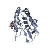 5bpbSC 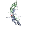 5bpuC  5bq8C 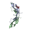 5bqbC 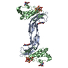 5bqcC 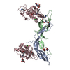 5bqeC S: Starting model for refinement C: citing same article ( |
|---|---|
| Similar structure data |
-Links
 PDBj
PDBj


- Assembly
| Deposited unit | 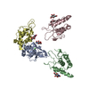
| ||||||||
|---|---|---|---|---|---|---|---|---|---|
| 1 | 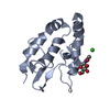
| ||||||||
| 2 | 
| ||||||||
| 3 | 
| ||||||||
| 4 | 
| ||||||||
| Unit cell |
|
-Components
| #1: Protein | / hFz4 / FzE4 Mass: 16705.232 Da / Num. of mol.: 4 / Fragment: cysteine-rich domain, UNP residues 42-179 Source method: isolated from a genetically manipulated source Source: (gene. exp.) Homo sapiens (human) / Gene: FZD4 / Plasmid: pHLsec-mVenus-12H / Cell line (production host): HEK293T / Production host: Homo sapiens (human) / References: UniProt: Q9ULV1#2: Sugar | ChemComp-NAG / N-Acetylglucosamine  Type: D-saccharide, beta linking / Mass: 221.208 Da / Num. of mol.: 4 / Fragment: cysteine-rich domain, UNP residues 42-179 Type: D-saccharide, beta linking / Mass: 221.208 Da / Num. of mol.: 4 / Fragment: cysteine-rich domain, UNP residues 42-179Source method: isolated from a genetically manipulated source Formula: C8H15NO6 / Source: (gene. exp.) Homo sapiens (human) / Gene: FZD4 / Plasmid: pHLsec-mVenus-12H / Cell line (production host): HEK293T / Production host: Homo sapiens (human)#3: Chemical | ChemComp-CL / | Chloride  Mass: 35.453 Da / Num. of mol.: 1 Mass: 35.453 Da / Num. of mol.: 1Source method: isolated from a genetically manipulated source Formula: Cl #4: Water | ChemComp-HOH / | Water Mass: 18.015 Da / Num. of mol.: 69 / Source method: isolated from a natural source / Formula: H2O Mass: 18.015 Da / Num. of mol.: 69 / Source method: isolated from a natural source / Formula: H2O |
|---|
-Experimental details
-Experiment
| Experiment | Method: X-RAY DIFFRACTION / Number of used crystals: 1 |
|---|
- Sample preparation
| Crystal | Density Matthews: 2.56 Å3/Da / Density % sol: 51.88 % |
|---|---|
| Crystal grow | Temperature: 294 K / Method: vapor diffusion, sitting drop Details: 0.1 M HEPES, pH 7.5, 0.1 M NaCl, 1.6 M ammonium sulfate |
-Data collection
| Diffraction | Mean temperature: 100 K | |||||||||||||||||||||||||||
|---|---|---|---|---|---|---|---|---|---|---|---|---|---|---|---|---|---|---|---|---|---|---|---|---|---|---|---|---|
| Diffraction source | Source: SYNCHROTRON / Site: Diamond / Beamline: I24 / Wavelength: 0.9686 Å | |||||||||||||||||||||||||||
| Detector | Type: DECTRIS PILATUS 6M / Detector: PIXEL / Date: Feb 17, 2013 | |||||||||||||||||||||||||||
| Radiation | Protocol: SINGLE WAVELENGTH / Monochromatic (M) / Laue (L): M / Scattering type: x-ray | |||||||||||||||||||||||||||
| Radiation wavelength | Wavelength: 0.9686 Å / Relative weight: 1 | |||||||||||||||||||||||||||
| Reflection | Resolution: 2.4→47.37 Å / Num. obs: 25975 / % possible obs: 99.5 % / Redundancy: 4 % / CC1/2: 0.999 / Rmerge(I) obs: 0.046 / Rpim(I) all: 0.026 / Net I/σ(I): 14.5 / Num. measured all: 103121 | |||||||||||||||||||||||||||
| Reflection shell | Diffraction-ID: 1 / Rejects: 0
|
-Phasing
| Phasing | Method: molecular replacement | |||||||||
|---|---|---|---|---|---|---|---|---|---|---|
| Phasing MR | Model details: Phaser MODE: MR_AUTO
|
- Processing
| Software |
| |||||||||||||||||||||||||||||||||||||||||||||||||||||||||||||||||||||||||||||||||||||||||||||||||||||||||||||||||||||||||||||
|---|---|---|---|---|---|---|---|---|---|---|---|---|---|---|---|---|---|---|---|---|---|---|---|---|---|---|---|---|---|---|---|---|---|---|---|---|---|---|---|---|---|---|---|---|---|---|---|---|---|---|---|---|---|---|---|---|---|---|---|---|---|---|---|---|---|---|---|---|---|---|---|---|---|---|---|---|---|---|---|---|---|---|---|---|---|---|---|---|---|---|---|---|---|---|---|---|---|---|---|---|---|---|---|---|---|---|---|---|---|---|---|---|---|---|---|---|---|---|---|---|---|---|---|---|---|---|
| Refinement | Method to determine structure: MOLECULAR REPLACEMENT Starting model: 5BPB Resolution: 2.4→65.88 Å / Cor.coef. Fo:Fc: 0.956 / Cor.coef. Fo:Fc free: 0.933 / SU B: 21.577 / SU ML: 0.218 / Cross valid method: THROUGHOUT / σ(F): 0 / ESU R Free: 0.235 / Stereochemistry target values: MAXIMUM LIKELIHOOD / Details: HYDROGENS HAVE BEEN ADDED IN THE RIDING POSITIONS
| |||||||||||||||||||||||||||||||||||||||||||||||||||||||||||||||||||||||||||||||||||||||||||||||||||||||||||||||||||||||||||||
| Solvent computation | Ion probe radii: 0.8 Å / Shrinkage radii: 0.8 Å / VDW probe radii: 1.2 Å / Solvent model: MASK | |||||||||||||||||||||||||||||||||||||||||||||||||||||||||||||||||||||||||||||||||||||||||||||||||||||||||||||||||||||||||||||
| Displacement parameters | Biso max: 160.44 Å2 / Biso mean: 74.887 Å2 / Biso min: 28.15 Å2
| |||||||||||||||||||||||||||||||||||||||||||||||||||||||||||||||||||||||||||||||||||||||||||||||||||||||||||||||||||||||||||||
| Refinement step | Cycle: final / Resolution: 2.4→65.88 Å
| |||||||||||||||||||||||||||||||||||||||||||||||||||||||||||||||||||||||||||||||||||||||||||||||||||||||||||||||||||||||||||||
| Refine LS restraints |
| |||||||||||||||||||||||||||||||||||||||||||||||||||||||||||||||||||||||||||||||||||||||||||||||||||||||||||||||||||||||||||||
| LS refinement shell | Resolution: 2.4→2.462 Å / Total num. of bins used: 20
| |||||||||||||||||||||||||||||||||||||||||||||||||||||||||||||||||||||||||||||||||||||||||||||||||||||||||||||||||||||||||||||
| Refinement TLS params. | Method: refined / Refine-ID: X-RAY DIFFRACTION
| |||||||||||||||||||||||||||||||||||||||||||||||||||||||||||||||||||||||||||||||||||||||||||||||||||||||||||||||||||||||||||||
| Refinement TLS group |
|
