 Movie
Movie Controller

[English] 日本語
 Yorodumi
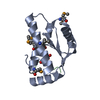
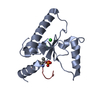
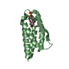
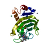
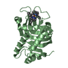
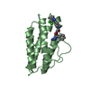
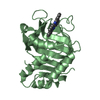
- PDB-6qtm: Crystal structure of the Sir4 H-BRCT domain in complex with Ty5 p... -
+ Open data

- Basic information
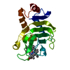
| Entry | Database: PDB / ID: 6qtm | ||||||
|---|---|---|---|---|---|---|---|
| Title | Crystal structure of the Sir4 H-BRCT domain in complex with Ty5 pS1095 peptide | ||||||
 Components Components |
| ||||||
 Keywords Keywords |  NUCLEAR PROTEIN / Heterochromatin NUCLEAR PROTEIN / Heterochromatin | ||||||
| Function / homology |  Function and homology information Function and homology informationtransposition / establishment of protein-containing complex localization to telomere / telomere tethering at nuclear periphery / chromatin silencing complex / silent mating-type cassette heterochromatin formation / positive regulation of heterochromatin formation / subtelomeric heterochromatin formation / nucleosome binding / heterochromatin formation / DNA integration ...transposition / establishment of protein-containing complex localization to telomere / telomere tethering at nuclear periphery / chromatin silencing complex / silent mating-type cassette heterochromatin formation / positive regulation of heterochromatin formation / subtelomeric heterochromatin formation / nucleosome binding / heterochromatin formation / DNA integration / double-strand break repair via nonhomologous end joining / RNA-directed DNA polymerase activity / RNA-DNA hybrid ribonuclease activity / double-stranded DNA binding / chromosome, telomeric region / molecular adaptor activity / DNA-directed DNA polymerase activity / RNA binding / nucleus / cytoplasmSimilarity search - Function | ||||||
| Biological species |  Saccharomyces cerevisiae S288C (yeast)Saccharomyces paradoxus (yeast) Saccharomyces cerevisiae S288C (yeast)Saccharomyces paradoxus (yeast) | ||||||
| Method | X-RAY DIFFRACTION / SYNCHROTRON / MOLECULAR REPLACEMENT / Resolution: 3 Å | ||||||
 Authors Authors | Gut, H. / Deshpande, I. / Keusch, J.J. / Challa, K. / Iesmantavicius, V. / Gasser, S.M. | ||||||
 Citation Citation | Journal: Embo J. / Year: 2019 Title: The Sir4 H-BRCT domain interacts with phospho-proteins to sequester and repress yeast heterochromatin. Authors: Deshpande, I. / Keusch, J.J. / Challa, K. / Iesmantavicius, V. / Gasser, S.M. / Gut, H. | ||||||
| History |
|
- Structure visualization
| Structure viewer | Molecule: MolmilJmol/JSmol |
|---|
- Downloads & links
-Download
| PDBx/mmCIF format | 6qtm.cif.gz | 158 KB | Display | PDBx/mmCIF format |
|---|---|---|---|---|
| PDB format | pdb6qtm.ent.gz | 132.6 KB | Display | PDB format |
| PDBx/mmJSON format | 6qtm.json.gz | Tree view | PDBx/mmJSON format | |
| Others |  Other downloads Other downloads |
-Validation report
| Arichive directory | https://data.pdbj.org/pub/pdb/validation_reports/qt/6qtmftp://data.pdbj.org/pub/pdb/validation_reports/qt/6qtm | HTTPS FTP |
|---|
-Related structure data


-Links
 PDBj
PDBj
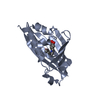
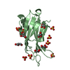
- Assembly
| Deposited unit | 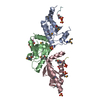
| ||||||||
|---|---|---|---|---|---|---|---|---|---|
| 1 | 
| ||||||||
| 2 | 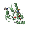
| ||||||||
| 3 | 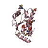
| ||||||||
| Unit cell |
|
-Components
| #1: Protein | Mass: 15059.491 Da / Num. of mol.: 3 Source method: isolated from a genetically manipulated source Details: Fragment, residues 961-1085 / Source: (gene. exp.) Saccharomyces cerevisiae S288C (yeast) / Gene: SIR4, ASD1, STE9, UTH2, YDR227W, YD9934.12 / Production host:  Escherichia coli BL21(DE3) (bacteria) / Variant (production host): B834 / References: UniProt: P11978 Escherichia coli BL21(DE3) (bacteria) / Variant (production host): B834 / References: UniProt: P11978#2: Protein/peptide | Mass: 1826.763 Da / Num. of mol.: 3 / Source method: obtained synthetically / Source: (synth.) Saccharomyces paradoxus (yeast) / References: UniProt: O42838, ribonuclease H#3: Chemical | ChemComp-SO4 / Sulfate  Mass: 96.063 Da / Num. of mol.: 6 / Source method: obtained synthetically / Formula: SO4 Mass: 96.063 Da / Num. of mol.: 6 / Source method: obtained synthetically / Formula: SO4#4: Water | ChemComp-HOH / | Water Mass: 18.015 Da / Num. of mol.: 7 / Source method: isolated from a natural source / Formula: H2O Mass: 18.015 Da / Num. of mol.: 7 / Source method: isolated from a natural source / Formula: H2O |
|---|
-Experimental details
-Experiment
| Experiment | Method: X-RAY DIFFRACTION / Number of used crystals: 1 |
|---|
- Sample preparation
| Crystal | Density Matthews: 2.34 Å3/Da / Density % sol: 47.52 % |
|---|---|
| Crystal grow | Temperature: 293 K / Method: vapor diffusion, hanging drop / pH: 5.6 Details: 2.0 M ammonium sulfate 0.2 M sodium potassium tartrate 0.1 M tri-sodium citrate pH 5.6 |
-Data collection
| Diffraction | Mean temperature: 100 K / Serial crystal experiment: N |
|---|---|
| Diffraction source | Source: SYNCHROTRON / Site: SLS  / Beamline: X06DA / Wavelength: 1 Å / Beamline: X06DA / Wavelength: 1 Å |
| Detector | Type: DECTRIS PILATUS 2M / Detector: PIXEL / Date: Jan 30, 2017 |
| Radiation | Protocol: SINGLE WAVELENGTH / Monochromatic (M) / Laue (L): M / Scattering type: x-ray |
| Radiation wavelength | Wavelength: 1 Å / Relative weight: 1 |
| Reflection | Resolution: 3→50 Å / Num. obs: 10009 / % possible obs: 97.1 % / Redundancy: 4.2 % / Biso Wilson estimate: 81.97 Å2 / CC1/2: 0.995 / Rsym value: 0.181 / Net I/σ(I): 7.7 |
| Reflection shell | Resolution: 3→3.08 Å / Mean I/σ(I) obs: 1.1 / CC1/2: 0.62 / % possible all: 95.8 |
- Processing
| Software |
| ||||||||||||||||||||||||||||||||||||||||||||||||||||||||||||||||||||||||||||||||||||||||||||||||||||||||||||||||||
|---|---|---|---|---|---|---|---|---|---|---|---|---|---|---|---|---|---|---|---|---|---|---|---|---|---|---|---|---|---|---|---|---|---|---|---|---|---|---|---|---|---|---|---|---|---|---|---|---|---|---|---|---|---|---|---|---|---|---|---|---|---|---|---|---|---|---|---|---|---|---|---|---|---|---|---|---|---|---|---|---|---|---|---|---|---|---|---|---|---|---|---|---|---|---|---|---|---|---|---|---|---|---|---|---|---|---|---|---|---|---|---|---|---|---|---|
| Refinement | Method to determine structure: MOLECULAR REPLACEMENT / Resolution: 3→49.33 Å / Cor.coef. Fo:Fc: 0.8691 / Cor.coef. Fo:Fc free: 0.8367 / Cross valid method: THROUGHOUT / σ(F): 0 / SU Rfree Blow DPI: 0.43
| ||||||||||||||||||||||||||||||||||||||||||||||||||||||||||||||||||||||||||||||||||||||||||||||||||||||||||||||||||
| Displacement parameters | Biso mean: 91.88 Å2
| ||||||||||||||||||||||||||||||||||||||||||||||||||||||||||||||||||||||||||||||||||||||||||||||||||||||||||||||||||
| Refine analyze | Luzzati coordinate error obs: 0.663 Å | ||||||||||||||||||||||||||||||||||||||||||||||||||||||||||||||||||||||||||||||||||||||||||||||||||||||||||||||||||
| Refinement step | Cycle: 1 / Resolution: 3→49.33 Å
| ||||||||||||||||||||||||||||||||||||||||||||||||||||||||||||||||||||||||||||||||||||||||||||||||||||||||||||||||||
| Refine LS restraints |
| ||||||||||||||||||||||||||||||||||||||||||||||||||||||||||||||||||||||||||||||||||||||||||||||||||||||||||||||||||
| LS refinement shell | Resolution: 3→3.35 Å / Total num. of bins used: 5
| ||||||||||||||||||||||||||||||||||||||||||||||||||||||||||||||||||||||||||||||||||||||||||||||||||||||||||||||||||
| Refinement TLS params. | Method: refined / Refine-ID: X-RAY DIFFRACTION
| ||||||||||||||||||||||||||||||||||||||||||||||||||||||||||||||||||||||||||||||||||||||||||||||||||||||||||||||||||
| Refinement TLS group |
|
