 Movie
Movie Controller

[English] 日本語
 Yorodumi
- PDB-5wrm: Mu2 subunit of the clathrin adaptor complex AP2 in complex with I... -
+ Open data

- Basic information
| Entry | Database: PDB / ID: 5wrm | ||||||
|---|---|---|---|---|---|---|---|
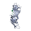
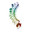
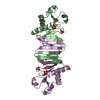
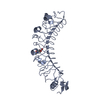
| Title | Mu2 subunit of the clathrin adaptor complex AP2 in complex with IRS-1 Y658 peptide | ||||||
 Components Components |
| ||||||
 Keywords Keywords |  ENDOCYTOSIS / clathrin adaptor AP-2 complex subunit / peptide complex ENDOCYTOSIS / clathrin adaptor AP-2 complex subunit / peptide complex | ||||||
| Function / homology |  Function and homology information Function and homology informationSOS-mediated signalling / IRS-related events triggered by IGF1R / cellular response to radiation / IRS-mediated signalling / IRS activation / PI3K/AKT activation / PI3K Cascade / PIP3 activates AKT signaling / Signaling by ALK / negative regulation of somatostatin secretion ...SOS-mediated signalling / IRS-related events triggered by IGF1R / cellular response to radiation / IRS-mediated signalling / IRS activation / PI3K/AKT activation / PI3K Cascade / PIP3 activates AKT signaling / Signaling by ALK / negative regulation of somatostatin secretion / positive regulation of glucagon secretion / epithelial cell migration / Gap junction degradation / Formation of annular gap junctions / WNT5A-dependent internalization of FZD2, FZD5 and ROR2 / LDL clearance / VLDLR internalisation and degradation / Retrograde neurotrophin signalling / Signal attenuation / WNT5A-dependent internalization of FZD4 / PI5P, PP2A and IER3 Regulate PI3K/AKT Signaling / extrinsic component of presynaptic endocytic zone membrane / MHC class II antigen presentation / AP-2 adaptor complex / regulation of vesicle size / postsynaptic neurotransmitter receptor internalization / Recycling pathway of L1 / Cargo recognition for clathrin-mediated endocytosis / positive regulation of synaptic vesicle endocytosis / Clathrin-mediated endocytosis / clathrin adaptor activity / positive regulation of fatty acid beta-oxidation / phosphatidylinositol 3-kinase activator activity / vesicle budding from membrane / clathrin-dependent endocytosis / mammary gland development / insulin receptor complex / positive regulation of glucose metabolic process / signal sequence binding / transmembrane receptor protein tyrosine kinase adaptor activity / cellular response to angiotensin / RAF/MAP kinase cascade / negative regulation of protein localization to plasma membrane / response to caffeine / positive regulation of mesenchymal cell proliferation / cellular response to fatty acid / low-density lipoprotein particle receptor binding / positive regulation of epithelial cell migration / Trafficking of GluR2-containing AMPA receptors / positive regulation of receptor internalization / synaptic vesicle endocytosis / protein localization to nucleus / negative regulation of insulin secretion / positive regulation of glycogen biosynthetic process / phosphatidylinositol 3-kinase binding / lipid catabolic process / positive regulation of insulin receptor signaling pathway / positive regulation of phosphorylation / clathrin-coated pit / cellular response to brain-derived neurotrophic factor stimulus / negative regulation of insulin receptor signaling pathway / insulin-like growth factor receptor binding / phosphotyrosine residue binding / SH2 domain binding / insulin-like growth factor receptor signaling pathway / ciliary basal body / protein kinase C binding / response to activity / phosphatidylinositol 3-kinase/protein kinase B signal transduction / caveola / positive regulation of glucose import / intracellular protein transport / response to insulin / insulin receptor binding / terminal bouton / receptor internalization / cytokine-mediated signaling pathway / cellular response to insulin stimulus / disordered domain specific binding / cell migration / signaling receptor complex adaptor activity / protein-macromolecule adaptor activity / insulin receptor signaling pathway / cytoplasmic vesicle / postsynapse / regulation of gene expression / protein-containing complex assembly / transmembrane transporter binding / positive regulation of phosphatidylinositol 3-kinase/protein kinase B signal transduction / protein domain specific binding / intracellular membrane-bounded organelle / synapse / glutamatergic synapse / lipid binding / protein-containing complex binding / protein kinase binding / nucleus / plasma membrane / cytosol / cytoplasmSimilarity search - Function | ||||||
| Biological species |  Rattus norvegicus (Norway rat) Rattus norvegicus (Norway rat) | ||||||
| Method | X-RAY DIFFRACTION / SYNCHROTRON / MOLECULAR REPLACEMENT / Resolution: 2.597 Å | ||||||
 Authors Authors | Yoneyama, Y. / Niwa, H. / Umehara, T. / Yokoyama, S. / Hakuno, F. / Takahashi, S. | ||||||
 Citation Citation | Journal: Elife / Year: 2018 Title: IRS-1 acts as an endocytic regulator of IGF-I receptor to facilitate sustained IGF signaling Authors: Yoneyama, Y. / Lanzerstorfer, P. / Niwa, H. / Umehara, T. / Shibano, T. / Yokoyama, S. / Chida, K. / Weghuber, J. / Hakuno, F. / Takahashi, S.I. | ||||||
| History |
|
- Structure visualization
| Structure viewer | Molecule: MolmilJmol/JSmol |
|---|
- Downloads & links
-Download
| PDBx/mmCIF format | 5wrm.cif.gz | 124.6 KB | Display | PDBx/mmCIF format |
|---|---|---|---|---|
| PDB format | pdb5wrm.ent.gz | 96.7 KB | Display | PDB format |
| PDBx/mmJSON format | 5wrm.json.gz | Tree view | PDBx/mmJSON format | |
| Others |  Other downloads Other downloads |
-Validation report
| Arichive directory | https://data.pdbj.org/pub/pdb/validation_reports/wr/5wrmftp://data.pdbj.org/pub/pdb/validation_reports/wr/5wrm | HTTPS FTP |
|---|
-Related structure data
| Related structure data | 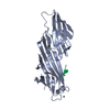 5wrkC 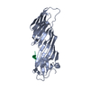 5wrlC 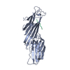 1bw8S C: citing same article ( S: Starting model for refinement |
|---|---|
| Similar structure data |


-Links
 PDBj
PDBj


- Assembly
| Deposited unit | 
| ||||||||
|---|---|---|---|---|---|---|---|---|---|
| 1 |
| ||||||||
| Unit cell |
| ||||||||
| Components on special symmetry positions |
|
-Components
| #1: Protein | Mass: 31798.293 Da / Num. of mol.: 1 / Fragment: UNP residues 158-435 Source method: isolated from a genetically manipulated source Source: (gene. exp.) Rattus norvegicus (Norway rat) / Gene: Ap2m1 / Production host:  Escherichia coli (E. coli) / References: UniProt: P84092 Escherichia coli (E. coli) / References: UniProt: P84092 |
|---|---|
| #2: Protein/peptide | IRS1 / IRS-1 / pp185 Mass: 903.099 Da / Num. of mol.: 1 / Fragment: UNP residues 657-664 / Source method: obtained synthetically / Source: (synth.) Rattus norvegicus (Norway rat) / References: UniProt: P35570 |
| #3: Water | ChemComp-HOH / Water Mass: 18.015 Da / Num. of mol.: 34 / Source method: isolated from a natural source / Formula: H2O Mass: 18.015 Da / Num. of mol.: 34 / Source method: isolated from a natural source / Formula: H2O |
-Experimental details
-Experiment
| Experiment | Method: X-RAY DIFFRACTION / Number of used crystals: 1 |
|---|
- Sample preparation
| Crystal | Density Matthews: 5.15 Å3/Da / Density % sol: 76.13 % Description: THE ENTRY CONTAINS FRIEDEL PAIRS IN F_PLUS/MINUS COLUMNS. |
|---|---|
| Crystal grow | Temperature: 291 K / Method: vapor diffusion, hanging drop Details: 2.3M NaCl, 0.4M Na/K Phosphate, 0.01M DTT, 15% Glycerol, 0.1M MES pH 6.5 |
-Data collection
| Diffraction | Mean temperature: 100 K |
|---|---|
| Diffraction source | Source: SYNCHROTRON / Site: SPring-8  / Beamline: BL26B2 / Wavelength: 1 Å / Beamline: BL26B2 / Wavelength: 1 Å |
| Detector | Type: RAYONIX MX225HE / Detector: CCD / Date: Jul 26, 2010 |
| Radiation | Protocol: SINGLE WAVELENGTH / Monochromatic (M) / Laue (L): M / Scattering type: x-ray |
| Radiation wavelength | Wavelength: 1 Å / Relative weight: 1 |
| Reflection | Resolution: 2.597→50 Å / Num. obs: 20659 / % possible obs: 100 % / Observed criterion σ(I): -3 / Redundancy: 11.4 % / Rsym value: 0.09 / Net I/σ(I): 26.5 |
| Reflection shell | Resolution: 2.6→2.64 Å / Redundancy: 11.5 % / Rmerge(I) obs: 1.977 / Mean I/σ(I) obs: 1.6 / CC1/2: 0.78 / % possible all: 100 |
- Processing
| Software |
| ||||||||||||||||||||||||||||||||||||||||||||||||||||||||||||||||||||||||||||||||||||||||||||||||||||||||||||||||||||||||||||||||||||||||||||||||||||||
|---|---|---|---|---|---|---|---|---|---|---|---|---|---|---|---|---|---|---|---|---|---|---|---|---|---|---|---|---|---|---|---|---|---|---|---|---|---|---|---|---|---|---|---|---|---|---|---|---|---|---|---|---|---|---|---|---|---|---|---|---|---|---|---|---|---|---|---|---|---|---|---|---|---|---|---|---|---|---|---|---|---|---|---|---|---|---|---|---|---|---|---|---|---|---|---|---|---|---|---|---|---|---|---|---|---|---|---|---|---|---|---|---|---|---|---|---|---|---|---|---|---|---|---|---|---|---|---|---|---|---|---|---|---|---|---|---|---|---|---|---|---|---|---|---|---|---|---|---|---|---|---|
| Refinement | Method to determine structure: MOLECULAR REPLACEMENT Starting model: 1BW8 Resolution: 2.597→35.927 Å / SU ML: 0.42 / Cross valid method: FREE R-VALUE / σ(F): 0.01 / Phase error: 29.28 Details: SF FILE CONTAINS FRIEDEL PAIRS UNDER I/F_MINUS AND I/F_PLUS COLUMNS.
| ||||||||||||||||||||||||||||||||||||||||||||||||||||||||||||||||||||||||||||||||||||||||||||||||||||||||||||||||||||||||||||||||||||||||||||||||||||||
| Solvent computation | Shrinkage radii: 0.9 Å / VDW probe radii: 1.11 Å | ||||||||||||||||||||||||||||||||||||||||||||||||||||||||||||||||||||||||||||||||||||||||||||||||||||||||||||||||||||||||||||||||||||||||||||||||||||||
| Refinement step | Cycle: LAST / Resolution: 2.597→35.927 Å
| ||||||||||||||||||||||||||||||||||||||||||||||||||||||||||||||||||||||||||||||||||||||||||||||||||||||||||||||||||||||||||||||||||||||||||||||||||||||
| Refine LS restraints |
| ||||||||||||||||||||||||||||||||||||||||||||||||||||||||||||||||||||||||||||||||||||||||||||||||||||||||||||||||||||||||||||||||||||||||||||||||||||||
| LS refinement shell |
| ||||||||||||||||||||||||||||||||||||||||||||||||||||||||||||||||||||||||||||||||||||||||||||||||||||||||||||||||||||||||||||||||||||||||||||||||||||||
| Refinement TLS params. | Method: refined / Refine-ID: X-RAY DIFFRACTION
| ||||||||||||||||||||||||||||||||||||||||||||||||||||||||||||||||||||||||||||||||||||||||||||||||||||||||||||||||||||||||||||||||||||||||||||||||||||||
| Refinement TLS group |
|
