[English] 日本語
Yorodumi
- PDB-2pr9: Mu2 adaptin subunit (AP50) of AP2 adaptor (second domain), comple... -

+
Open data


ID or keywords:

Loading...

-
Basic information

Entry
Database: PDB / ID: 2pr9
TitleMu2 adaptin subunit (AP50) of AP2 adaptor (second domain), complexed with GABAA receptor-gamma2 subunit-derived internalization peptide DEEYGYECL
Components
  • AP-2 complex subunit mu-1
  • GABA(A) receptor subunit gamma-2 peptide
KeywordsENDOCYTOSIS / adaptor / internalization peptide complex / inhibitory neurotransmitter receptor
Function / homology
Function and homology information


GABA receptor activation / Gap junction degradation / Formation of annular gap junctions / WNT5A-dependent internalization of FZD2, FZD5 and ROR2 / LDL clearance / VLDLR internalisation and degradation / Retrograde neurotrophin signalling / WNT5A-dependent internalization of FZD4 / extrinsic component of presynaptic endocytic zone membrane / MHC class II antigen presentation ...GABA receptor activation / Gap junction degradation / Formation of annular gap junctions / WNT5A-dependent internalization of FZD2, FZD5 and ROR2 / LDL clearance / VLDLR internalisation and degradation / Retrograde neurotrophin signalling / WNT5A-dependent internalization of FZD4 / extrinsic component of presynaptic endocytic zone membrane / MHC class II antigen presentation / AP-2 adaptor complex / inhibitory synapse / regulation of vesicle size / postsynaptic neurotransmitter receptor internalization / GABA receptor complex / inhibitory extracellular ligand-gated monoatomic ion channel activity / Recycling pathway of L1 / GABA-gated chloride ion channel activity / cellular response to histamine / inhibitory synapse assembly / GABA-A receptor activity / GABA-A receptor complex / Cargo recognition for clathrin-mediated endocytosis / positive regulation of synaptic vesicle endocytosis / Clathrin-mediated endocytosis / clathrin adaptor activity / vesicle budding from membrane / clathrin-dependent endocytosis / signal sequence binding / postsynaptic specialization membrane / synaptic transmission, GABAergic / gamma-aminobutyric acid signaling pathway / negative regulation of protein localization to plasma membrane / chloride transport / low-density lipoprotein particle receptor binding / chloride channel activity / adult behavior / Trafficking of GluR2-containing AMPA receptors / chloride channel complex / positive regulation of receptor internalization / synaptic vesicle endocytosis / GABA-ergic synapse / regulation of postsynaptic membrane potential / clathrin-coated pit / chloride transmembrane transport / dendrite membrane / post-embryonic development / transmitter-gated monoatomic ion channel activity involved in regulation of postsynaptic membrane potential / intracellular protein transport / cytoplasmic vesicle membrane / terminal bouton / receptor internalization / disordered domain specific binding / cytoplasmic vesicle / chemical synaptic transmission / postsynapse / protein-containing complex assembly / transmembrane transporter binding / neuron projection / axon / synapse / glutamatergic synapse / lipid binding / plasma membrane
Similarity search - Function
Mu homology domain, subdomain B / Mu2, C-terminal domain / AP-2 complex subunit mu, N-terminal / Gamma-aminobutyric-acid A receptor, gamma 2 subunit / Gamma-aminobutyric acid receptor subunit gamma-1/4 / Clathrin adaptor complexes medium chain signature 1. / Clathrin adaptor, mu subunit / Clathrin adaptor, mu subunit, conserved site / Clathrin adaptor complexes medium chain signature 2. / Adaptor complexes medium subunit family ...Mu homology domain, subdomain B / Mu2, C-terminal domain / AP-2 complex subunit mu, N-terminal / Gamma-aminobutyric-acid A receptor, gamma 2 subunit / Gamma-aminobutyric acid receptor subunit gamma-1/4 / Clathrin adaptor complexes medium chain signature 1. / Clathrin adaptor, mu subunit / Clathrin adaptor, mu subunit, conserved site / Clathrin adaptor complexes medium chain signature 2. / Adaptor complexes medium subunit family / AP complex, mu/sigma subunit / Clathrin adaptor complex small chain / AP-2 complex subunit mu, C-terminal superfamily / Mu homology domain / Mu homology domain (MHD) profile. / Longin-like domain superfamily / Gamma-aminobutyric acid A receptor/Glycine receptor alpha / Neurotransmitter-gated ion-channel, conserved site / Neurotransmitter-gated ion-channels signature. / Neurotransmitter-gated ion-channel transmembrane domain / Neurotransmitter-gated ion-channel transmembrane region / Neurotransmitter-gated ion-channel transmembrane domain superfamily / Neuronal acetylcholine receptor / Neurotransmitter-gated ion-channel / Neurotransmitter-gated ion-channel ligand-binding domain / Neurotransmitter-gated ion-channel ligand-binding domain superfamily / Neurotransmitter-gated ion-channel ligand binding domain / Immunoglobulin-like / Sandwich / Mainly Beta
Similarity search - Domain/homology
Gamma-aminobutyric acid receptor subunit gamma-2 / AP-2 complex subunit mu
Similarity search - Component
Biological speciesRattus norvegicus (Norway rat)
MethodX-RAY DIFFRACTION / SYNCHROTRON / MOLECULAR REPLACEMENT / Resolution: 2.51 Å
AuthorsVahedi-Faridi, A. / Haucke, V. / Kittler, J.T. / Kukhtina, V. / Moss, S.J. / Saenger, W. / Chen, G.-J. / Tretter, V. / Smith, K. / Yan, Z. ...Vahedi-Faridi, A. / Haucke, V. / Kittler, J.T. / Kukhtina, V. / Moss, S.J. / Saenger, W. / Chen, G.-J. / Tretter, V. / Smith, K. / Yan, Z. / McAinsh, K. / Arancibia-Carcamo, L.
CitationJournal: Proc.Natl.Acad.Sci.Usa / Year: 2008
Title: Regulation of synaptic inhibition by phospho-dependent binding of the AP2 complex to a YECL motif in the GABAA receptor gamma2 subunit.
Authors: Kittler, J.T. / Chen, G. / Kukhtina, V. / Vahedi-Faridi, A. / Gu, Z. / Tretter, V. / Smith, K.R. / McAinsh, K. / Arancibia-Carcamo, I.L. / Saenger, W. / Haucke, V. / Yan, Z. / Moss, S.J.
History
DepositionMay 4, 2007Deposition site: RCSB / Processing site: RCSB
Revision 1.0Mar 18, 2008Provider: repository / Type: Initial release
Revision 1.1Jul 13, 2011Group: Advisory / Version format compliance
Revision 1.2Aug 30, 2023Group: Data collection / Database references / Refinement description
Category: chem_comp_atom / chem_comp_bond ...chem_comp_atom / chem_comp_bond / database_2 / pdbx_initial_refinement_model / struct_ref_seq_dif
Item: _database_2.pdbx_DOI / _database_2.pdbx_database_accession / _struct_ref_seq_dif.details
Remark 300BIOMOLECULE THIS ENTRY CONTAINS THE CRYSTALLOGRAPHIC ASYMMETRIC UNIT WHICH CONSISTS OF 2 CHAIN(S). ...BIOMOLECULE THIS ENTRY CONTAINS THE CRYSTALLOGRAPHIC ASYMMETRIC UNIT WHICH CONSISTS OF 2 CHAIN(S). AUTHORS STATE THAT THE BIOLOGICAL MOLECULE FOR THE PROTEIN IS UNKNOWN.

-
Structure visualization

Structure viewerMolecule:
MolmilJmol/JSmol

Downloads & links

-
Assembly

Deposited unit
A: AP-2 complex subunit mu-1
P: GABA(A) receptor subunit gamma-2 peptide


Theoretical massNumber of molelcules
Total (without water)35,3362
Polymers35,3362
Non-polymers00
Water1,06359
1


  • Idetical with deposited unit
  • defined by software
TypeNameSymmetry operationNumber
identity operation1_555x,y,z1
Buried area1060 Å2
ΔGint-5.2 kcal/mol
Surface area14880 Å2
MethodPISA
Unit cell
Length a, b, c (Å)126.300, 126.300, 74.650
Angle α, β, γ (deg.)90.000, 90.000, 120.000
Int Tables number172
Space group name H-MP64

-
Components

#1: Protein AP-2 complex subunit mu-1


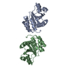
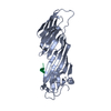
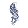
Mass: 34100.840 Da / Num. of mol.: 1 / Fragment: second domain (residues 158-435)
Source method: isolated from a genetically manipulated source
Source: (gene. exp.) Rattus norvegicus (Norway rat) / Gene: Ap2m1 / Plasmid: pET-28a(+) / Species (production host): Escherichia coli / Production host: Escherichia coli BL21(DE3) (bacteria) / Strain (production host): BL21 (DE3) / References: UniProt: P84092
#2: Protein/peptide GABA(A) receptor subunit gamma-2 peptide


Mass: 1235.232 Da / Num. of mol.: 1 / Source method: obtained synthetically
Details: The peptide was chemically synthesized. This sequence occurs naturally in mouse and rat.
References: UniProt: P18508
#3: Water ChemComp-HOH / water / Water


Mass: 18.015 Da / Num. of mol.: 59 / Source method: isolated from a natural source / Formula: H2O

-
Experimental details

-
Experiment

ExperimentMethod: X-RAY DIFFRACTION / Number of used crystals: 1

-
Sample preparation

CrystalDensity Matthews: 4.86 Å3/Da / Density % sol: 74.7 %
Crystal growTemperature: 291 K / Method: vapor diffusion, sitting drop / pH: 6
Details: 2.0 M sodium formate, 0.1 M Na-acetate, pH 6.0, VAPOR DIFFUSION, SITTING DROP, temperature 291K

-
Data collection

DiffractionMean temperature: 100 K
Diffraction sourceSource: SYNCHROTRON / Site: BESSY / Beamline: 14.2 / Wavelength: 0.95373
DetectorType: MAR CCD 165 mm / Detector: CCD / Date: Mar 29, 2006 / Details: mirrors
RadiationMonochromator: GRAPHITE / Protocol: SINGLE WAVELENGTH / Monochromatic (M) / Laue (L): M / Scattering type: x-ray
Radiation wavelengthWavelength: 0.95373 Å / Relative weight: 1
ReflectionResolution: 2.5→50 Å / Num. obs: 22817 / % possible obs: 96.6 % / Observed criterion σ(I): -3 / Redundancy: 5.3 % / Biso Wilson estimate: 66.389 Å2 / Rmerge(I) obs: 0.064 / Net I/σ(I): 20.14
Reflection shellResolution: 2.5→2.75 Å / Redundancy: 2.1 % / Rmerge(I) obs: 0.659 / Mean I/σ(I) obs: 1 / Num. unique all: 3053 / % possible all: 68.9

-
Phasing

Phasing MR
Highest resolutionLowest resolution
Rotation3.18 Å9.97 Å
Translation3.18 Å9.97 Å

-
Processing

Software
NameVersionClassificationNB
XSCALEdata scaling
MOLREPphasing
REFMACrefinement
PDB_EXTRACT2data extraction
ADSCQuantumdata collection
XDSdata reduction
RefinementMethod to determine structure: MOLECULAR REPLACEMENT
Starting model: PDB ENTRY 1BW8
Resolution: 2.51→9.97 Å / Cor.coef. Fo:Fc: 0.953 / Cor.coef. Fo:Fc free: 0.932 / SU B: 15.237 / SU ML: 0.16 / TLS residual ADP flag: LIKELY RESIDUAL / Cross valid method: THROUGHOUT / σ(F): 0 / ESU R: 0.209 / ESU R Free: 0.193 / Stereochemistry target values: MAXIMUM LIKELIHOOD / Details: HYDROGENS HAVE BEEN ADDED IN THE RIDING POSITIONS
RfactorNum. reflection% reflectionSelection details
Rfree0.24 913 4 %RANDOM
Rwork0.203 ---
obs0.204 22817 100 %-
Solvent computationIon probe radii: 0.8 Å / Shrinkage radii: 0.8 Å / VDW probe radii: 1.4 Å / Solvent model: MASK
Displacement parametersBiso mean: 52.027 Å2
Baniso -1Baniso -2Baniso -3
1--0.98 Å2-0.49 Å20 Å2
2---0.98 Å20 Å2
3---1.46 Å2
Refinement stepCycle: LAST / Resolution: 2.51→9.97 Å
ProteinNucleic acidLigandSolventTotal
Num. atoms2115 0 0 59 2174
Refine LS restraints
Refine-IDTypeDev idealDev ideal targetNumber
X-RAY DIFFRACTIONr_bond_refined_d0.0360.0222157
X-RAY DIFFRACTIONr_angle_refined_deg2.6411.9812905
X-RAY DIFFRACTIONr_dihedral_angle_1_deg9.4335260
X-RAY DIFFRACTIONr_dihedral_angle_2_deg38.85823.22290
X-RAY DIFFRACTIONr_dihedral_angle_3_deg20.16915409
X-RAY DIFFRACTIONr_dihedral_angle_4_deg18.5131517
X-RAY DIFFRACTIONr_chiral_restr0.1930.2321
X-RAY DIFFRACTIONr_gen_planes_refined0.010.021585
X-RAY DIFFRACTIONr_nbd_refined0.2410.2815
X-RAY DIFFRACTIONr_nbtor_refined0.3260.21431
X-RAY DIFFRACTIONr_xyhbond_nbd_refined0.1720.290
X-RAY DIFFRACTIONr_symmetry_vdw_refined0.1240.223
X-RAY DIFFRACTIONr_symmetry_hbond_refined0.3470.211
X-RAY DIFFRACTIONr_mcbond_it1.4221.51370
X-RAY DIFFRACTIONr_mcangle_it2.26422143
X-RAY DIFFRACTIONr_scbond_it3.6173934
X-RAY DIFFRACTIONr_scangle_it5.3734.5762
LS refinement shellResolution: 2.512→2.573 Å / Total num. of bins used: 20
RfactorNum. reflection% reflection
Rfree0.455 63 -
Rwork0.431 1504 -
obs-1567 100 %
Refinement TLS params.

Method: refined / Refine-ID: X-RAY DIFFRACTION

IDL112)L122)L132)L222)L232)L332)S11 (Å °)S12 (Å °)S13 (Å °)S21 (Å °)S22 (Å °)S23 (Å °)S31 (Å °)S32 (Å °)S33 (Å °)T112)T122)T132)T222)T232)T332)Origin x (Å)Origin y (Å)Origin z (Å)
15.48291.2208-2.55231.8875-0.03522.1263-0.28310.0438-0.5692-0.16670.0883-0.20080.2371-0.1060.1949-0.04710.0397-0.0645-0.11440.0668-0.1159-43.838240.619925.3328
241.338-12.4676-16.44330.32722.762521.4441-1.4882-3.13160.63072.80121.19680.8733-0.2527-0.75720.29140.0735-0.0263-0.01160.0299-0.06180.0273-32.244550.928527.7458
Refinement TLS group

Refine-ID: X-RAY DIFFRACTION / Selection: ALL

IDRefine TLS-IDAuth asym-IDLabel asym-IDAuth seq-IDLabel seq-ID
11AA159 - 21923 - 83
21AA238 - 255102 - 119
31AA261 - 435125 - 299
42PB1 - 101 - 10

+
About Yorodumi

-
News

-
Feb 9, 2022. New format data for meta-information of EMDB entries

New format data for meta-information of EMDB entries

  • Version 3 of the EMDB header file is now the official format.
  • The previous official version 1.9 will be removed from the archive.

Related info.:EMDB header

External links:wwPDB to switch to version 3 of the EMDB data model

-
Aug 12, 2020. Covid-19 info

Covid-19 info

URL: https://pdbj.org/emnavi/covid19.php

New page: Covid-19 featured information page in EM Navigator.

Related info.:Covid-19 info / Mar 5, 2020. Novel coronavirus structure data

+
Mar 5, 2020. Novel coronavirus structure data

Novel coronavirus structure data

Related info.:Yorodumi Speices / Aug 12, 2020. Covid-19 info

External links:COVID-19 featured content - PDBj / Molecule of the Month (242):Coronavirus Proteases

+
Jan 31, 2019. EMDB accession codes are about to change! (news from PDBe EMDB page)

EMDB accession codes are about to change! (news from PDBe EMDB page)

  • The allocation of 4 digits for EMDB accession codes will soon come to an end. Whilst these codes will remain in use, new EMDB accession codes will include an additional digit and will expand incrementally as the available range of codes is exhausted. The current 4-digit format prefixed with “EMD-” (i.e. EMD-XXXX) will advance to a 5-digit format (i.e. EMD-XXXXX), and so on. It is currently estimated that the 4-digit codes will be depleted around Spring 2019, at which point the 5-digit format will come into force.
  • The EM Navigator/Yorodumi systems omit the EMD- prefix.

Related info.:Q: What is EMD? / ID/Accession-code notation in Yorodumi/EM Navigator

External links:EMDB Accession Codes are Changing Soon! / Contact to PDBj

+
Jul 12, 2017. Major update of PDB

Major update of PDB

  • wwPDB released updated PDB data conforming to the new PDBx/mmCIF dictionary.
  • This is a major update changing the version number from 4 to 5, and with Remediation, in which all the entries are updated.
  • In this update, many items about electron microscopy experimental information are reorganized (e.g. em_software).
  • Now, EM Navigator and Yorodumi are based on the updated data.

External links:wwPDB Remediation / Enriched Model Files Conforming to OneDep Data Standards Now Available in the PDB FTP Archive

-
Yorodumi

Thousand views of thousand structures

  • Yorodumi is a browser for structure data from EMDB, PDB, SASBDB, etc.
  • This page is also the successor to EM Navigator detail page, and also detail information page/front-end page for Omokage search.
  • The word "yorodu" (or yorozu) is an old Japanese word meaning "ten thousand". "mi" (miru) is to see.

Related info.:EMDB / PDB / SASBDB / Comparison of 3 databanks / Yorodumi Search / Aug 31, 2016. New EM Navigator & Yorodumi / Yorodumi Papers / Jmol/JSmol / Function and homology information / Changes in new EM Navigator and Yorodumi

Read more