 Movie
Movie Controller


+ Open data

- Basic information
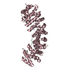
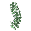
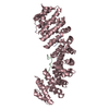
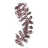
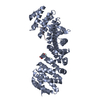
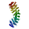
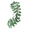
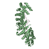
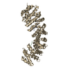
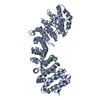
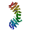
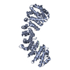
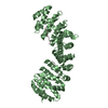
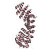
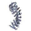
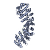
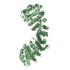
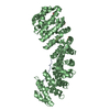
| Entry | Database: PDB / ID: 3l3q | ||||||
|---|---|---|---|---|---|---|---|
| Title | Mouse importin alpha-pepTM NLS peptide complex | ||||||
 Components Components |
| ||||||
 Keywords Keywords |  PROTEIN TRANSPORT / ARMADILLO REPEATS PROTEIN TRANSPORT / ARMADILLO REPEATS | ||||||
| Function / homology |  Function and homology information Function and homology informationSensing of DNA Double Strand Breaks / entry of viral genome into host nucleus through nuclear pore complex via importin / positive regulation of viral life cycle / NLS-dependent protein nuclear import complex / postsynapse to nucleus signaling pathway / nuclear import signal receptor activity / nuclear localization sequence binding / NLS-bearing protein import into nucleus / host cell / cytoplasmic stress granule ...Sensing of DNA Double Strand Breaks / entry of viral genome into host nucleus through nuclear pore complex via importin / positive regulation of viral life cycle / NLS-dependent protein nuclear import complex / postsynapse to nucleus signaling pathway / nuclear import signal receptor activity / nuclear localization sequence binding / NLS-bearing protein import into nucleus / host cell / cytoplasmic stress granule / protein import into nucleus / DNA-binding transcription factor binding / postsynaptic density / glutamatergic synapse / nucleoplasm / nucleus / cytosolSimilarity search - Function | ||||||
| Biological species |  Mus musculus (house mouse) Mus musculus (house mouse) | ||||||
| Method | X-RAY DIFFRACTION / SYNCHROTRON / MOLECULAR REPLACEMENT / Resolution: 2.3 Å | ||||||
 Authors Authors | Takeda, A.A.S. / Kobe, B. / Fontes, M.R.M. | ||||||
 Citation Citation | Journal: J.Biol.Chem. / Year: 2010 Title: Probing the specificity of binding to the major nuclear localization sequence-binding site of importin-alpha using oriented peptide library screening. Authors: Yang, S.N.Y. / Takeda, A.A.S. / Fontes, M.R.M. / Harris, J.M. / Jans, D.A. / Kobe, B. | ||||||
| History |
|
- Structure visualization
| Structure viewer | Molecule: MolmilJmol/JSmol |
|---|
- Downloads & links
-Download
| PDBx/mmCIF format | 3l3q.cif.gz | 101.7 KB | Display | PDBx/mmCIF format |
|---|---|---|---|---|
| PDB format | pdb3l3q.ent.gz | 77.6 KB | Display | PDB format |
| PDBx/mmJSON format | 3l3q.json.gz | Tree view | PDBx/mmJSON format | |
| Others |  Other downloads Other downloads |
-Validation report
| Arichive directory | https://data.pdbj.org/pub/pdb/validation_reports/l3/3l3qftp://data.pdbj.org/pub/pdb/validation_reports/l3/3l3q | HTTPS FTP |
|---|
-Related structure data
| Related structure data |  1pjnS S: Starting model for refinement |
|---|---|
| Similar structure data |


-Links
 PDBj
PDBj


- Assembly
| Deposited unit | 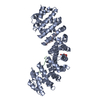
| ||||||||
|---|---|---|---|---|---|---|---|---|---|
| 1 |
| ||||||||
| Unit cell |
|
-Components
| #1: Protein | / Importin alpha-2 / Karyopherin subunit alpha-2 / SRP1-alpha / RAG cohort protein 1 / Pendulin / ...Importin alpha-2 / Karyopherin subunit alpha-2 / SRP1-alpha / RAG cohort protein 1 / Pendulin / Pore targeting complex 58 kDa subunit / PTAC58 / Importin alpha P1 Mass: 46271.953 Da / Num. of mol.: 1 / Fragment: NLS binding domain (UNP residues 71-497) Source method: isolated from a genetically manipulated source Source: (gene. exp.) Mus musculus (house mouse) / Gene: Kpna2, Rch1 / Plasmid: pET30a / Production host:  Escherichia coli (E. coli) / Strain (production host): BL21(DE3) pLysS / References: UniProt: P52293 Escherichia coli (E. coli) / Strain (production host): BL21(DE3) pLysS / References: UniProt: P52293 | ||||
|---|---|---|---|---|---|
| #2: Protein/peptide | Mass: 1801.031 Da / Num. of mol.: 2 / Source method: obtained synthetically Details: This sequence was obtained by an oriented peptide library approach to probe the specificity of the major NLS binding site in importin alpha. #3: Chemical | ChemComp-FLC / | Citric acid  Mass: 189.100 Da / Num. of mol.: 1 / Source method: obtained synthetically / Formula: C6H5O7 Mass: 189.100 Da / Num. of mol.: 1 / Source method: obtained synthetically / Formula: C6H5O7#4: Water | ChemComp-HOH / | Water Mass: 18.015 Da / Num. of mol.: 107 / Source method: isolated from a natural source / Formula: H2O Mass: 18.015 Da / Num. of mol.: 107 / Source method: isolated from a natural source / Formula: H2O |
-Experimental details
-Experiment
| Experiment | Method: X-RAY DIFFRACTION / Number of used crystals: 1 |
|---|
- Sample preparation
| Crystal | Density Matthews: 3.62 Å3/Da / Density % sol: 66.05 % |
|---|---|
| Crystal grow | Temperature: 293 K / Method: vapor diffusion, hanging drop / pH: 6 Details: Sodium Citrate, DTT, pH 6, VAPOR DIFFUSION, HANGING DROP, temperature 293K |
-Data collection
| Diffraction | Mean temperature: 100 K |
|---|---|
| Diffraction source | Source: SYNCHROTRON / Site: LNLS  / Beamline: W01B-MX2 / Wavelength: 1.55 Å / Beamline: W01B-MX2 / Wavelength: 1.55 Å |
| Detector | Type: MARMOSAIC 225 mm CCD / Detector: CCD / Date: Mar 13, 2008 |
| Radiation | Monochromator: SAGITALLY FOCUSED Si(111) / Protocol: SINGLE WAVELENGTH / Monochromatic (M) / Laue (L): M / Scattering type: x-ray |
| Radiation wavelength | Wavelength: 1.55 Å / Relative weight: 1 |
| Reflection | Resolution: 2.3→40 Å / Num. all: 29414 / Num. obs: 29414 / % possible obs: 89.6 % / Observed criterion σ(F): 0 / Observed criterion σ(I): -3 / Redundancy: 3.9 % / Rmerge(I) obs: 0.094 |
| Reflection shell | Resolution: 2.3→2.38 Å / Redundancy: 4.4 % / Rmerge(I) obs: 0.094 / % possible all: 89.3 |
- Processing
| Software |
| ||||||||||||||||||||||||||||||||||||||||||||||||||||||||||||||||||||||||||||||||||||||||||||||||||||||||||||||||||||||||||||||||||||||||||||||||||||||||||||||||||||||||||
|---|---|---|---|---|---|---|---|---|---|---|---|---|---|---|---|---|---|---|---|---|---|---|---|---|---|---|---|---|---|---|---|---|---|---|---|---|---|---|---|---|---|---|---|---|---|---|---|---|---|---|---|---|---|---|---|---|---|---|---|---|---|---|---|---|---|---|---|---|---|---|---|---|---|---|---|---|---|---|---|---|---|---|---|---|---|---|---|---|---|---|---|---|---|---|---|---|---|---|---|---|---|---|---|---|---|---|---|---|---|---|---|---|---|---|---|---|---|---|---|---|---|---|---|---|---|---|---|---|---|---|---|---|---|---|---|---|---|---|---|---|---|---|---|---|---|---|---|---|---|---|---|---|---|---|---|---|---|---|---|---|---|---|---|---|---|---|---|---|---|---|---|
| Refinement | Method to determine structure: MOLECULAR REPLACEMENT Starting model: PDB ENTRY 1PJN Resolution: 2.3→33.79 Å / Cor.coef. Fo:Fc: 0.953 / Cor.coef. Fo:Fc free: 0.937 / SU B: 4.905 / SU ML: 0.121 / Cross valid method: THROUGHOUT / ESU R: 0.229 / ESU R Free: 0.195 / Stereochemistry target values: MAXIMUM LIKELIHOOD
| ||||||||||||||||||||||||||||||||||||||||||||||||||||||||||||||||||||||||||||||||||||||||||||||||||||||||||||||||||||||||||||||||||||||||||||||||||||||||||||||||||||||||||
| Solvent computation | Ion probe radii: 0.8 Å / Shrinkage radii: 0.8 Å / VDW probe radii: 1.2 Å / Solvent model: MASK | ||||||||||||||||||||||||||||||||||||||||||||||||||||||||||||||||||||||||||||||||||||||||||||||||||||||||||||||||||||||||||||||||||||||||||||||||||||||||||||||||||||||||||
| Displacement parameters | Biso mean: 52.563 Å2
| ||||||||||||||||||||||||||||||||||||||||||||||||||||||||||||||||||||||||||||||||||||||||||||||||||||||||||||||||||||||||||||||||||||||||||||||||||||||||||||||||||||||||||
| Refinement step | Cycle: LAST / Resolution: 2.3→33.79 Å
| ||||||||||||||||||||||||||||||||||||||||||||||||||||||||||||||||||||||||||||||||||||||||||||||||||||||||||||||||||||||||||||||||||||||||||||||||||||||||||||||||||||||||||
| Refine LS restraints |
| ||||||||||||||||||||||||||||||||||||||||||||||||||||||||||||||||||||||||||||||||||||||||||||||||||||||||||||||||||||||||||||||||||||||||||||||||||||||||||||||||||||||||||
| LS refinement shell | Resolution: 2.298→2.357 Å / Total num. of bins used: 20
|
