 Movie
Movie Controller

[English] 日本語
 Yorodumi
- PDB-2kqt: Solid-state NMR structure of the M2 transmembrane peptide of the ... -
+ Open data

- Basic information
| Entry | Database: PDB / ID: 2kqt | ||||||
|---|---|---|---|---|---|---|---|
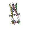
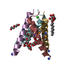
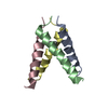
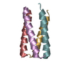
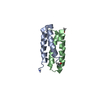
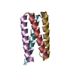
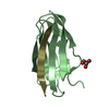
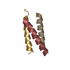
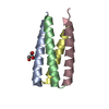
| Title | Solid-state NMR structure of the M2 transmembrane peptide of the influenza A virus in DMPC lipid bilayers bound to deuterated amantadine | ||||||
 Components Components | M2 protein M2 proton channel M2 proton channel | ||||||
 Keywords Keywords | TRANSPORT PROTEIN / influenza / transmembrane / amantadine / REDOR | ||||||
| Function / homology |  Function and homology information Function and homology informationsuppression by virus of host autophagy / proton transmembrane transporter activity / : / protein complex oligomerization / monoatomic ion channel activity / host cell plasma membrane / virion membrane / membrane / identical protein bindingSimilarity search - Function | ||||||
| Method | SOLID-STATE NMR / simulated annealing, Monte Carlo | ||||||
| Model details | fewest violations, model 1 | ||||||
 Authors Authors | Cady, S.D. / Schmidt-Rohr, K. / Wang, J. / Soto, C.S. / DeGrado, W.F. / Hong, M. | ||||||
 Citation Citation | Journal: Biophys.J. / Year: 2007 Title: Backbone structure of the amantadine-blocked trans-membrane domain M2 proton channel from Influenza A virus. Authors: Hu, J. / Asbury, T. / Achuthan, S. / Li, C. / Bertram, R. / Quine, J.R. / Fu, R. / Cross, T.A. | ||||||
| History |
| ||||||
| Remark 0 | THIS ENTRY 2KQT REFLECTS AN ALTERNATIVE MODELING OF THE ORIGINAL STRUCTURAL DATA 2H95 and 2KAD |
- Structure visualization
| Structure viewer | Molecule: MolmilJmol/JSmol |
|---|
- Downloads & links
-Download
| PDBx/mmCIF format | 2kqt.cif.gz | 535.2 KB | Display | PDBx/mmCIF format |
|---|---|---|---|---|
| PDB format | pdb2kqt.ent.gz | 451.7 KB | Display | PDB format |
| PDBx/mmJSON format | 2kqt.json.gz | Tree view | PDBx/mmJSON format | |
| Others |  Other downloads Other downloads |
-Validation report
| Arichive directory | https://data.pdbj.org/pub/pdb/validation_reports/kq/2kqtftp://data.pdbj.org/pub/pdb/validation_reports/kq/2kqt | HTTPS FTP |
|---|
-Related structure data
| Related structure data | |
|---|---|
| Similar structure data | |
| Other databases |


-Links
 PDBj
PDBj- Assembly
| Deposited unit | 
| |||||||||
|---|---|---|---|---|---|---|---|---|---|---|
| 1 |
| |||||||||
| NMR ensembles |
|
-Components
| #1: Protein/peptide | M2 proton channel Mass: 2730.295 Da / Num. of mol.: 4 / Fragment: residues 22-46 / Source method: obtained synthetically / Details: Solid-phase synthesis / References: UniProt: Q9YP62, UniProt: O70632*PLUS #2: Chemical | ChemComp-308 / ( | Amantadine  Mass: 151.249 Da / Num. of mol.: 1 / Source method: obtained synthetically / Formula: C10H17N / Comment: medication, antagonist*YM Mass: 151.249 Da / Num. of mol.: 1 / Source method: obtained synthetically / Formula: C10H17N / Comment: medication, antagonist*YM |
|---|
-Experimental details
-Experiment
| Experiment | Method: SOLID-STATE NMR | ||||||||||||||||||||||||||||||||||||||||||||||||||||||||||||||||||||
|---|---|---|---|---|---|---|---|---|---|---|---|---|---|---|---|---|---|---|---|---|---|---|---|---|---|---|---|---|---|---|---|---|---|---|---|---|---|---|---|---|---|---|---|---|---|---|---|---|---|---|---|---|---|---|---|---|---|---|---|---|---|---|---|---|---|---|---|---|---|
| NMR experiment |
|
- Sample preparation
| Details |
| |||||||||||||||||||||||||||||||||||||||||||||||||||||||||||||||||||||||||||||||||||||||||||||||||||||||||||||||||||||||||||||||||||||
|---|---|---|---|---|---|---|---|---|---|---|---|---|---|---|---|---|---|---|---|---|---|---|---|---|---|---|---|---|---|---|---|---|---|---|---|---|---|---|---|---|---|---|---|---|---|---|---|---|---|---|---|---|---|---|---|---|---|---|---|---|---|---|---|---|---|---|---|---|---|---|---|---|---|---|---|---|---|---|---|---|---|---|---|---|---|---|---|---|---|---|---|---|---|---|---|---|---|---|---|---|---|---|---|---|---|---|---|---|---|---|---|---|---|---|---|---|---|---|---|---|---|---|---|---|---|---|---|---|---|---|---|---|---|---|
| Sample |
|