 Movie
Movie Controller

[English] 日本語
 Yorodumi
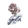
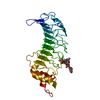
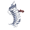
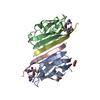
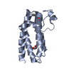
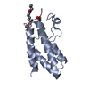
- PDB-2bp3: Crystal structure of Filamin A domain 17 and GPIb alpha cytoplasm... -
+ Open data

- Basic information
| Entry | Database: PDB / ID: 2bp3 | ||||||
|---|---|---|---|---|---|---|---|
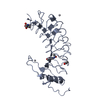
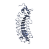
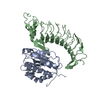
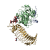
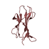
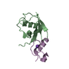
| Title | Crystal structure of Filamin A domain 17 and GPIb alpha cytoplasmic domain complex | ||||||
 Components Components |
| ||||||
 Keywords Keywords |  STRUCTURAL PROTEIN / CYTOSKELETON-COMPLEX / ACTIN BINDING PROTEIN / CYTOSKELETON / COMPLEX STRUCTURAL PROTEIN / CYTOSKELETON-COMPLEX / ACTIN BINDING PROTEIN / CYTOSKELETON / COMPLEX | ||||||
| Function / homology |  Function and homology information Function and homology informationEnhanced binding of GP1BA variant to VWF multimer:collagen / Defective binding of VWF variant to GPIb:IX:V / thrombin-activated receptor activity / regulation of membrane repolarization during atrial cardiac muscle cell action potential / regulation of membrane repolarization during cardiac muscle cell action potential / establishment of Sertoli cell barrier / Myb complex / formation of radial glial scaffolds / glycoprotein Ib-IX-V complex / adenylate cyclase-inhibiting dopamine receptor signaling pathway ...Enhanced binding of GP1BA variant to VWF multimer:collagen / Defective binding of VWF variant to GPIb:IX:V / thrombin-activated receptor activity / regulation of membrane repolarization during atrial cardiac muscle cell action potential / regulation of membrane repolarization during cardiac muscle cell action potential / establishment of Sertoli cell barrier / Myb complex / formation of radial glial scaffolds / glycoprotein Ib-IX-V complex / adenylate cyclase-inhibiting dopamine receptor signaling pathway / positive regulation of integrin-mediated signaling pathway / cytoplasmic sequestering of protein / positive regulation of leukocyte tethering or rolling / tubulin deacetylation / actin crosslink formation / blood coagulation, intrinsic pathway / protein localization to bicellular tight junction / OAS antiviral response / positive regulation of actin filament bundle assembly / positive regulation of neuron migration / positive regulation of potassium ion transmembrane transport / Cell-extracellular matrix interactions / Defective F9 activation / early endosome to late endosome transport / Platelet Adhesion to exposed collagen / apical dendrite / positive regulation of neural precursor cell proliferation / cell-cell junction organization / positive regulation of platelet activation / protein localization to cell surface / Fc-gamma receptor I complex binding / negative regulation of transcription by RNA polymerase I / wound healing, spreading of cells / megakaryocyte development / GP1b-IX-V activation signalling / cortical cytoskeleton / positive regulation of axon regeneration / receptor clustering / SMAD binding / regulation of blood coagulation / RHO GTPases activate PAKs / actin filament bundle / Platelet Aggregation (Plug Formation) / brush border / semaphorin-plexin signaling pathway / cilium assembly / mitotic spindle assembly / potassium channel regulator activity / epithelial to mesenchymal transition / blood vessel remodeling / axonal growth cone / heart morphogenesis / positive regulation of substrate adhesion-dependent cell spreading / regulation of cell migration / release of sequestered calcium ion into cytosol / fibrinolysis / Intrinsic Pathway of Fibrin Clot Formation / extracellular matrix / dendritic shaft / protein kinase C binding / protein localization to plasma membrane / G protein-coupled receptor binding / actin filament / synapse organization / RUNX1 regulates genes involved in megakaryocyte differentiation and platelet function / mRNA transcription by RNA polymerase II / trans-Golgi network / establishment of protein localization / cell morphogenesis / negative regulation of DNA-binding transcription factor activity / negative regulation of protein catabolic process / cerebral cortex development / platelet activation / Z disc / small GTPase binding / platelet aggregation / kinase binding / positive regulation of protein import into nucleus / actin filament binding / cell-cell junction / blood coagulation / actin cytoskeleton / Platelet degranulation / negative regulation of neuron projection development / GTPase binding / perikaryon / actin cytoskeleton organization / postsynapse / angiogenesis / positive regulation of canonical NF-kappaB signal transduction / DNA-binding transcription factor binding / transmembrane transporter binding / cell surface receptor signaling pathway / protein stabilization / cell adhesion / cadherin binding / external side of plasma membrane / focal adhesion / glutamatergic synapse / nucleolusSimilarity search - Function | ||||||
| Biological species |  HOMO SAPIENS (human) HOMO SAPIENS (human) | ||||||
| Method | X-RAY DIFFRACTION / SYNCHROTRON / MOLECULAR REPLACEMENT / Resolution: 2.32 Å | ||||||
 Authors Authors | Pudas, R. / Ylanne, J. | ||||||
 Citation Citation | Journal: Blood / Year: 2006 Title: The Structure of the Gpib-Filamin a Complex. Authors: Nakamura, F. / Pudas, R. / Heikkinen, O. / Permi, P. / Kilpelainen, I. / Munday, A.D. / Hartwig, J.H. / Stossel, T.P. / Ylanne, J. | ||||||
| History |
| ||||||
| Remark 700 | SHEET THE SHEET STRUCTURE OF THIS MOLECULE IS BIFURCATED. IN ORDER TO REPRESENT THIS FEATURE IN ... SHEET THE SHEET STRUCTURE OF THIS MOLECULE IS BIFURCATED. IN ORDER TO REPRESENT THIS FEATURE IN THE SHEET RECORDS BELOW, TWO SHEETS ARE DEFINED. |
- Structure visualization
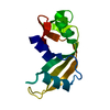
| Structure viewer | Molecule: MolmilJmol/JSmol |
|---|
- Downloads & links
-Download
| PDBx/mmCIF format | 2bp3.cif.gz | 54.2 KB | Display | PDBx/mmCIF format |
|---|---|---|---|---|
| PDB format | pdb2bp3.ent.gz | 38.2 KB | Display | PDB format |
| PDBx/mmJSON format | 2bp3.json.gz | Tree view | PDBx/mmJSON format | |
| Others |  Other downloads Other downloads |
-Validation report
| Arichive directory | https://data.pdbj.org/pub/pdb/validation_reports/bp/2bp3ftp://data.pdbj.org/pub/pdb/validation_reports/bp/2bp3 | HTTPS FTP |
|---|
-Related structure data
| Related structure data |  2aavC  1v05S S: Starting model for refinement C: citing same article ( |
|---|---|
| Similar structure data |


-Links
 PDBj
PDBj


- Assembly
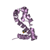
| Deposited unit | 
| ||||||||
|---|---|---|---|---|---|---|---|---|---|
| 1 | 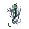
| ||||||||
| 2 | 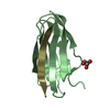
| ||||||||
| Unit cell |
|
-Components
| #1: Protein | FLNA / ALPHA-FILAMIN / FILAMIN 1 / ENDOTHELIAL ACTIN-BINDING PROTEIN / ACTIN-BINDING PROTEIN 280 / ABP-280 ...ALPHA-FILAMIN / FILAMIN 1 / ENDOTHELIAL ACTIN-BINDING PROTEIN / ACTIN-BINDING PROTEIN 280 / ABP-280 / NONMUSCLE FILAMIN Mass: 9976.017 Da / Num. of mol.: 2 / Fragment: ROD DOMAIN, RESIDUES 1863-1956 Source method: isolated from a genetically manipulated source Source: (gene. exp.) HOMO SAPIENS (human) / Plasmid: PGEX4T-3 / Production host:  ESCHERICHIA COLI (E. coli) / Strain (production host): BL21 / References: UniProt: P21333 ESCHERICHIA COLI (E. coli) / Strain (production host): BL21 / References: UniProt: P21333#2: Protein/peptide | Mass: 2563.014 Da / Num. of mol.: 2 / Fragment: CYTOPLASMIC DOMAIN, RESIDUES 572-593 / Source method: obtained synthetically / Source: (synth.) HOMO SAPIENS (human) / References: UniProt: P07359#3: Chemical | Glycerol  Mass: 92.094 Da / Num. of mol.: 2 / Source method: obtained synthetically / Formula: C3H8O3 Mass: 92.094 Da / Num. of mol.: 2 / Source method: obtained synthetically / Formula: C3H8O3#4: Water | ChemComp-HOH / | Water Mass: 18.015 Da / Num. of mol.: 28 / Source method: isolated from a natural source / Formula: H2O Mass: 18.015 Da / Num. of mol.: 28 / Source method: isolated from a natural source / Formula: H2OSequence details | THE THREE N-TERMINAL RESIDUES OF CHAINS A AND B ARE FROM THE EXPRESSION | |
|---|
-Experimental details
-Experiment
| Experiment | Method: X-RAY DIFFRACTION / Number of used crystals: 1 |
|---|
- Sample preparation
| Crystal | Density Matthews: 3.62 Å3/Da / Density % sol: 65.78 % |
|---|---|
| Crystal grow | pH: 8.2 Details: 1.75M AMMONIUM PHOSPHATE, PH 8.2, AFTER MICROSEEDING: 1.25M AMMONIUM SULFATE PH 8.2 |
-Data collection
| Diffraction | Mean temperature: 100 K |
|---|---|
| Diffraction source | Source: SYNCHROTRON / Site: ESRF  / Beamline: ID23-1 / Wavelength: 0.9795 / Beamline: ID23-1 / Wavelength: 0.9795 |
| Detector | Type: MARRESEARCH / Detector: CCD / Date: Dec 5, 2004 / Details: TOROIDAL MIRROR |
| Radiation | Monochromator: SILICON (1 1 1) CHANNEL-CUT / Protocol: SINGLE WAVELENGTH / Monochromatic (M) / Laue (L): M / Scattering type: x-ray |
| Radiation wavelength | Wavelength: 0.9795 Å / Relative weight: 1 |
| Reflection | Resolution: 2.32→19.88 Å / Num. obs: 12876 / % possible obs: 86.9 % / Redundancy: 6 % / Rmerge(I) obs: 0.09 / Net I/σ(I): 14 |
| Reflection shell | Resolution: 2.31→2.45 Å / Redundancy: 3.7 % / Rmerge(I) obs: 0.53 / Mean I/σ(I) obs: 3.13 / % possible all: 56.7 |
- Processing
| Software |
| ||||||||||||||||||||||||||||||||||||||||||||||||||||||||||||||||||||||||||||||||||||||||||||||||||||||||||||||||||||||||||||||||||||||||||||||||||||||||||||||||||||||||||||||||||||||
|---|---|---|---|---|---|---|---|---|---|---|---|---|---|---|---|---|---|---|---|---|---|---|---|---|---|---|---|---|---|---|---|---|---|---|---|---|---|---|---|---|---|---|---|---|---|---|---|---|---|---|---|---|---|---|---|---|---|---|---|---|---|---|---|---|---|---|---|---|---|---|---|---|---|---|---|---|---|---|---|---|---|---|---|---|---|---|---|---|---|---|---|---|---|---|---|---|---|---|---|---|---|---|---|---|---|---|---|---|---|---|---|---|---|---|---|---|---|---|---|---|---|---|---|---|---|---|---|---|---|---|---|---|---|---|---|---|---|---|---|---|---|---|---|---|---|---|---|---|---|---|---|---|---|---|---|---|---|---|---|---|---|---|---|---|---|---|---|---|---|---|---|---|---|---|---|---|---|---|---|---|---|---|---|
| Refinement | Method to determine structure: MOLECULAR REPLACEMENT Starting model: PDB ENTRY 1V05 Resolution: 2.32→19.88 Å / Cor.coef. Fo:Fc: 0.94 / Cor.coef. Fo:Fc free: 0.92 / SU B: 5.978 / SU ML: 0.147 / Cross valid method: THROUGHOUT / ESU R: 0.263 / ESU R Free: 0.217 / Stereochemistry target values: MAXIMUM LIKELIHOOD Details: HYDROGENS HAVE BEEN ADDED IN THE RIDING POSITIONS. FLEXIBLE SIDE CHAINS OF SOME RESIDUES LOCATED IN THE LOOP REGIONS COULD NOT BE BUILD USING AVAILABLE DENSITY AND THESE RESIDUES WERE ...Details: HYDROGENS HAVE BEEN ADDED IN THE RIDING POSITIONS. FLEXIBLE SIDE CHAINS OF SOME RESIDUES LOCATED IN THE LOOP REGIONS COULD NOT BE BUILD USING AVAILABLE DENSITY AND THESE RESIDUES WERE MODELED: A 1881 ASN, A 1882 LYS, A 1892 ASP, A 1916 GLN, B 1891 LYS, B 1892 ASP, B 1909 GLU, B 1916 GLN, B 1917 ASP
| ||||||||||||||||||||||||||||||||||||||||||||||||||||||||||||||||||||||||||||||||||||||||||||||||||||||||||||||||||||||||||||||||||||||||||||||||||||||||||||||||||||||||||||||||||||||
| Solvent computation | Ion probe radii: 0.8 Å / Shrinkage radii: 0.8 Å / VDW probe radii: 1.2 Å / Solvent model: BABINET MODEL WITH MASK | ||||||||||||||||||||||||||||||||||||||||||||||||||||||||||||||||||||||||||||||||||||||||||||||||||||||||||||||||||||||||||||||||||||||||||||||||||||||||||||||||||||||||||||||||||||||
| Displacement parameters | Biso mean: 57.94 Å2
| ||||||||||||||||||||||||||||||||||||||||||||||||||||||||||||||||||||||||||||||||||||||||||||||||||||||||||||||||||||||||||||||||||||||||||||||||||||||||||||||||||||||||||||||||||||||
| Refinement step | Cycle: LAST / Resolution: 2.32→19.88 Å
| ||||||||||||||||||||||||||||||||||||||||||||||||||||||||||||||||||||||||||||||||||||||||||||||||||||||||||||||||||||||||||||||||||||||||||||||||||||||||||||||||||||||||||||||||||||||
| Refine LS restraints |
|