 Movie
Movie Controller

[English] 日本語
 Yorodumi
- PDB-1nyj: The closed state structure of M2 protein H+ channel by solid stat... -
+ Open data

- Basic information
| Entry | Database: PDB / ID: 1nyj | ||||||
|---|---|---|---|---|---|---|---|
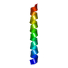
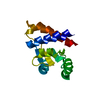
| Title | The closed state structure of M2 protein H+ channel by solid state NMR spectroscopy | ||||||
 Components Components | Matrix protein M2 | ||||||
 Keywords Keywords |  VIRAL PROTEIN / influenza A virus / membrane protein structure / M2 proton channel / solid state NMR VIRAL PROTEIN / influenza A virus / membrane protein structure / M2 proton channel / solid state NMR | ||||||
| Function / homology |  Function and homology information Function and homology informationsuppression by virus of host autophagy / proton transmembrane transporter activity / : / protein complex oligomerization / monoatomic ion channel activity / host cell plasma membrane / virion membrane / membraneSimilarity search - Function | ||||||
| Method | SOLID-STATE NMR / simulated annealing | ||||||
 Authors Authors | Nishimura, K. / Kim, S. / Zhang, L. / Cross, T.A. | ||||||
 Citation Citation | Journal: Biochemistry / Year: 2002 Title: The closed state of a H+ channel helical bundle combining precise orientational and distance restraints from solid state NMR Authors: Nishimura, K. / Kim, S. / Zhang, L. / Cross, T.A. #1: Journal: Protein Sci. / Year: 2001Title: Structure of the transmembrane region of the M2 protein H+ channel Authors: Wang, J. / Kim, S. / Kovacs, F. / Cross, T.A. | ||||||
| History |
|
- Structure visualization
| Structure viewer | Molecule: MolmilJmol/JSmol |
|---|
- Downloads & links
-Download
| PDBx/mmCIF format | 1nyj.cif.gz | 23.4 KB | Display | PDBx/mmCIF format |
|---|---|---|---|---|
| PDB format | pdb1nyj.ent.gz | 16.4 KB | Display | PDB format |
| PDBx/mmJSON format | 1nyj.json.gz | Tree view | PDBx/mmJSON format | |
| Others |  Other downloads Other downloads |
-Validation report
| Arichive directory | https://data.pdbj.org/pub/pdb/validation_reports/ny/1nyjftp://data.pdbj.org/pub/pdb/validation_reports/ny/1nyj | HTTPS FTP |
|---|
-Related structure data
| Related structure data | |
|---|---|
| Similar structure data |


-Links
 PDBj
PDBj- Assembly
| Deposited unit | 
| |||||||||
|---|---|---|---|---|---|---|---|---|---|---|
| 1 |
| |||||||||
| NMR ensembles |
|
-Components
| #1: Protein/peptide | Mass: 2730.295 Da / Num. of mol.: 4 / Fragment: Transmembrane peptide (residue 22-46) / Source method: obtained synthetically Details: THE PEPTIDE WAS SYNTHESIZED USING SOLID PHASE PEPTIDE SYNTHESIS. THIS SEQUENCE OCCURS NATURALLY IN THE INFLUENZA A VIRUS (UDORN/72). References: UniProt: P35938 |
|---|
-Experimental details
-Experiment
| Experiment | Method: SOLID-STATE NMR |
|---|---|
| NMR experiment | Type: Solid state NMR PISEMA and REDOR |
| NMR details | Text: The heteronuclear distance was obtained by means of solid state NMR REDOR experiments. 13C-REDOR was performed on a DMX-300 with an XY8-pulse sequence for irradiation of 15N nuclei to ...Text: The heteronuclear distance was obtained by means of solid state NMR REDOR experiments. 13C-REDOR was performed on a DMX-300 with an XY8-pulse sequence for irradiation of 15N nuclei to compensate for errors in the flip angle, off resonance effect, and variation in the H1 field. The spinning speed was controlled at 4000 1 Hz and the experiments were performed at 38 C. REDOR and full echo spectra were recorded at various dipolar evolution times, Nctr, (where Nc and tr are the rotor cycle number and rotor period, respectively) from 2 to 16 ms to observe reasonable dipolar dephasing of the signals. |
- Sample preparation
| Details | Contents: To prepare an unoriented hydrated lipid bilayer sample, M2-TMP and dimyristoylphosphatidylcholine (DMPC) in a 1:16 molar ratio were co-solubilized in trifluoroethanol (TFE). After ...Contents: To prepare an unoriented hydrated lipid bilayer sample, M2-TMP and dimyristoylphosphatidylcholine (DMPC) in a 1:16 molar ratio were co-solubilized in trifluoroethanol (TFE). After lyophilization, the white powder was hydrated by adding 50% (by total sample dry weight) pH 7.0, HPLC grade water followed by incubation for 2 days at 42 oC. The sample was then dispersed in 30 ml of 20 mM Citric/Na2HPO4 buffer at pH 7.0 and incubated at 45 oC for 2 hours before centrifuging at 20000 x g for 3 hours. After excess water was removed, the pellet was transferred to an eppendorf tube, tightly sealed to maintain the same hydration level, and then incubated at 45 oC for 2 days. Then, the sample was transferred to a glass insert for a Bruker 7 mm spinner and sealed with epoxy. Solvent system: To prepare an oriented bilayer sample, 20 mg of M2-TMP and 38.5 mg of DMPC (1:8 molar ratio) were co-dissolved in 1.5 ml of TFE. The sample was spread on 50 glass slides. After drying ...Solvent system: To prepare an oriented bilayer sample, 20 mg of M2-TMP and 38.5 mg of DMPC (1:8 molar ratio) were co-dissolved in 1.5 ml of TFE. The sample was spread on 50 glass slides. After drying the organic solvent from the slides approximately 1.2 l of buffer (0.2 M phosphate and 0.1 M citric acid, pH 7.0) was added to each slide and then they were stacked into a glass tubing with a 6.0 x 6.0 mm internal dimension cross section. |
|---|---|
| Sample conditions | Ionic strength: none / pH: 7.0 / Pressure: ambient / Temperature: 303.00 K |
| Crystal grow | *PLUS Method: other / Details: NMR |
-NMR measurement
| Radiation | Protocol: SINGLE WAVELENGTH / Monochromatic (M) / Laue (L): M | |||||||||||||||
|---|---|---|---|---|---|---|---|---|---|---|---|---|---|---|---|---|
| Radiation wavelength | Relative weight: 1 | |||||||||||||||
| NMR spectrometer |
|
- Processing
| NMR software | Name: TORC / Version: V5.4 / Developer: Ketchem, Roux, Cross / Classification: refinement |
|---|---|
| Refinement | Method: simulated annealing / Software ordinal: 1 Details: The symmetric, tetrameric bundle model of M2-TMP was constructed from the monomer structure (1MP6). The M2-TMP monomer coordinates were obtained by a geometrical search using a search ...Details: The symmetric, tetrameric bundle model of M2-TMP was constructed from the monomer structure (1MP6). The M2-TMP monomer coordinates were obtained by a geometrical search using a search algorithim to obtain a minimum of the global penalty function that incorporates all the orientational restraints and the CHARMM empirical function. The orientational restraints imposed on the structure during refinement are 15 15N chemical shifts and 15 15N-1H dipolar couplings from PISEMA experiments. The refinement was carried out in vacuo with the initial coordinates of an ideal a-helix structure (3.6 residues per turn) having a range of tilt and rotational orientations with respect to the bilayer spanning the values obtained from the PISA wheels. The resulting tetrameric bundle model was used to search the side chain orientations in accord with the experimentally measured distance between 15ND1 His37 and 13CG Trp41. Both chi 1 and chi 2 angles of the residues were searched extensively using 10 increments to discern whether the interaction was intramolecular or intermolecular and to find out which residues accounted for the observed spin interaction before characterizing the rotameric states of the sidechains. Note that while the His37 and Trp41 sidechain rotameric states are defined by the distance restraint, the rotameric states of other residues are taken from the backbone dependent sidechain rotamer library. |
| NMR ensemble | Conformer selection criteria: The tetrameric oligomer conformation of M2-TMP was constructed using the monomer structure refined by solid-state NMR orientational data. The monomer structure was the ...Conformer selection criteria: The tetrameric oligomer conformation of M2-TMP was constructed using the monomer structure refined by solid-state NMR orientational data. The monomer structure was the lowest energy conformer from 30 simulated annealing attempts. Conformers calculated total number: 1 / Conformers submitted total number: 1 |
