 Movie
Movie Controller

[English] 日本語
 Yorodumi
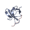
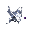
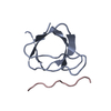
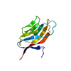
- PDB-2j6o: ATYPICAL POLYPROLINE RECOGNITION BY THE CMS N-TERMINAL SH3 DOMAIN... -
+ Open data

- Basic information
| Entry | Database: PDB / ID: 2j6o | ||||||
|---|---|---|---|---|---|---|---|
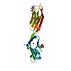
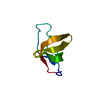
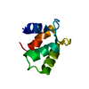
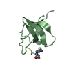
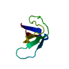
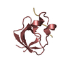
| Title | ATYPICAL POLYPROLINE RECOGNITION BY THE CMS N-TERMINAL SH3 DOMAIN. CMS:CD2 HETEROTRIMER | ||||||
 Components Components |
| ||||||
 Keywords Keywords |  PROTEIN BINDING / PHOSPHORYLATION / ADAPTOR PROTEIN / EGFR DOWNREGULATION / CMS / SH3 DOMAIN / SH3-BINDING / COILED COIL / SH3 DOMAIN RECOGNITION / SIGNALING PROTEIN PROTEIN BINDING / PHOSPHORYLATION / ADAPTOR PROTEIN / EGFR DOWNREGULATION / CMS / SH3 DOMAIN / SH3-BINDING / COILED COIL / SH3 DOMAIN RECOGNITION / SIGNALING PROTEIN | ||||||
| Function / homology |  Function and homology information Function and homology informationpositive regulation of myeloid dendritic cell activation / response to glial cell derived neurotrophic factor / membrane raft polarization / negative regulation of small GTPase mediated signal transduction / transforming growth factor beta1 production / localization of cell / Rab protein signal transduction / negative regulation of transforming growth factor beta1 production / slit diaphragm / response to transforming growth factor beta ...positive regulation of myeloid dendritic cell activation / response to glial cell derived neurotrophic factor / membrane raft polarization / negative regulation of small GTPase mediated signal transduction / transforming growth factor beta1 production / localization of cell / Rab protein signal transduction / negative regulation of transforming growth factor beta1 production / slit diaphragm / response to transforming growth factor beta / podocyte differentiation / endothelium development / immunological synapse formation / nerve growth factor signaling pathway / cell-cell adhesion mediated by cadherin / collateral sprouting / protein heterooligomerization / renal albumin absorption / substrate-dependent cell migration, cell extension / membrane organization / phosphatidylinositol 3-kinase regulatory subunit binding / natural killer cell mediated cytotoxicity / cell-cell junction organization / filopodium assembly / natural killer cell activation / podosome / Nephrin family interactions / heterotypic cell-cell adhesion / regulation of T cell differentiation / clathrin binding / maintenance of blood-brain barrier / nuclear envelope lumen / glucose import / cell leading edge / filamentous actin / neurotrophin TRK receptor signaling pathway / centriolar satellite / protein secretion / lymph node development / adipose tissue development / stress-activated MAPK cascade / ruffle / ERK1 and ERK2 cascade / T cell activation / actin filament polymerization / phosphatidylinositol 3-kinase/protein kinase B signal transduction / trans-Golgi network membrane / liver development / positive regulation of interleukin-8 production / actin filament organization / positive regulation of protein secretion / regulation of actin cytoskeleton organization / Cell surface interactions at the vascular wall / synapse organization / response to virus / regulation of synaptic plasticity / protein catabolic process / response to insulin / neuromuscular junction / lipid metabolic process / structural constituent of cytoskeleton / cytoplasmic side of plasma membrane / receptor tyrosine kinase binding / fibrillar center / cell-cell adhesion / SH3 domain binding / response to wounding / positive regulation of protein localization to nucleus / : / male gonad development / positive regulation of tumor necrosis factor production / positive regulation of type II interferon production / actin filament binding / cell-cell junction / cell migration / actin cytoskeleton / late endosome / signaling receptor activity / T cell receptor signaling pathway / growth cone / protein-containing complex assembly / response to oxidative stress / vesicle / negative regulation of neuron apoptotic process / cell population proliferation / cell surface receptor signaling pathway / cadherin binding / inflammatory response / cell cycle / external side of plasma membrane / axon / cell division / signaling receptor binding / apoptotic process / dendrite / Golgi apparatus / cell surface / signal transduction / protein-containing complex / extracellular exosomeSimilarity search - Function | ||||||
| Biological species |  HOMO SAPIENS (human) HOMO SAPIENS (human) | ||||||
| Method | X-RAY DIFFRACTION / MOLECULAR REPLACEMENT / Resolution: 2.23 Å | ||||||
 Authors Authors | Moncalian, G. / Cardenes, N. / Deribe, Y.L. / Spinola-Amilibia, M. / Dikic, I. / Bravo, J. | ||||||
 Citation Citation | Journal: J.Biol.Chem. / Year: 2006 Title: Atypical Polyproline Recognition by the Cms N-Terminal SH3 Domain. Authors: Moncalian, G. / Cardenes, N. / Deribe, Y.L. / Spinola-Amilibia, M. / Dikic, I. / Bravo, J. | ||||||
| History |
|
- Structure visualization
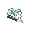
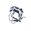
| Structure viewer | Molecule: MolmilJmol/JSmol |
|---|
- Downloads & links
-Download
| PDBx/mmCIF format | 2j6o.cif.gz | 27.2 KB | Display | PDBx/mmCIF format |
|---|---|---|---|---|
| PDB format | pdb2j6o.ent.gz | 18.1 KB | Display | PDB format |
| PDBx/mmJSON format | 2j6o.json.gz | Tree view | PDBx/mmJSON format | |
| Others |  Other downloads Other downloads |
-Validation report
| Arichive directory | https://data.pdbj.org/pub/pdb/validation_reports/j6/2j6oftp://data.pdbj.org/pub/pdb/validation_reports/j6/2j6o | HTTPS FTP |
|---|
-Related structure data


-Links
 PDBj
PDBj


- Assembly
| Deposited unit | 
| ||||||||
|---|---|---|---|---|---|---|---|---|---|
| 1 |
| ||||||||
| Unit cell |
| ||||||||
| Components on special symmetry positions |
|
-Components
| #1: Protein | Mass: 7412.285 Da / Num. of mol.: 1 / Fragment: SH3 DOMAIN, RESIDUES 1-62 Source method: isolated from a genetically manipulated source Source: (gene. exp.) HOMO SAPIENS (human) / Plasmid: PET21 / Production host:  ESCHERICHIA COLI (E. coli) / Strain (production host): BL21(DE3) / References: UniProt: Q9Y5K6 ESCHERICHIA COLI (E. coli) / Strain (production host): BL21(DE3) / References: UniProt: Q9Y5K6 |
|---|---|
| #2: Protein/peptide | Mass: 1119.382 Da / Num. of mol.: 1 / Fragment: CMS BINDING SEQUENCE, RESIDUES 324-333 / Source method: obtained synthetically / Source: (synth.) HOMO SAPIENS (human) / References: UniProt: P06729 |
| #3: Water | ChemComp-HOH / Water Mass: 18.015 Da / Num. of mol.: 46 / Source method: isolated from a natural source / Formula: H2O Mass: 18.015 Da / Num. of mol.: 46 / Source method: isolated from a natural source / Formula: H2O |
-Experimental details
-Experiment
| Experiment | Method: X-RAY DIFFRACTION / Number of used crystals: 1 |
|---|
- Sample preparation
| Crystal | Density Matthews: 2.34 Å3/Da / Density % sol: 47.42 % |
|---|---|
| Crystal grow | pH: 5.5 / Details: 20% PEG3000, 0.1M ACETATE PH 5.5 |
-Data collection
| Diffraction | Mean temperature: 110 K |
|---|---|
| Diffraction source | Source: ROTATING ANODE / Type: ENRAF-NONIUS FR591 / Wavelength: 1.54179 |
| Detector | Type: MARRESEARCH / Detector: CCD / Date: Jun 7, 2004 / Details: MIRRORS |
| Radiation | Protocol: SINGLE WAVELENGTH / Monochromatic (M) / Laue (L): M / Scattering type: x-ray |
| Radiation wavelength | Wavelength: 1.54179 Å / Relative weight: 1 |
| Reflection | Resolution: 2.22→34.36 Å / Num. obs: 3933 / % possible obs: 98 % / Observed criterion σ(I): 0 / Redundancy: 16.66 % / Biso Wilson estimate: 33 Å2 / Rmerge(I) obs: 0.11 / Net I/σ(I): 25.02 |
| Reflection shell | Resolution: 2.22→2.26 Å / Redundancy: 14.22 % / Rmerge(I) obs: 0.41 / Mean I/σ(I) obs: 15.74 / % possible all: 86.6 |
- Processing
| Software |
| ||||||||||||||||||||||||||||||||||||||||||||||||||||||||||||||||||||||||||||||||||||||||||||||||||||||||||||||||||||||||||||||||||||||||||||||||||||||||||||||||||||||||||||||||||||||
|---|---|---|---|---|---|---|---|---|---|---|---|---|---|---|---|---|---|---|---|---|---|---|---|---|---|---|---|---|---|---|---|---|---|---|---|---|---|---|---|---|---|---|---|---|---|---|---|---|---|---|---|---|---|---|---|---|---|---|---|---|---|---|---|---|---|---|---|---|---|---|---|---|---|---|---|---|---|---|---|---|---|---|---|---|---|---|---|---|---|---|---|---|---|---|---|---|---|---|---|---|---|---|---|---|---|---|---|---|---|---|---|---|---|---|---|---|---|---|---|---|---|---|---|---|---|---|---|---|---|---|---|---|---|---|---|---|---|---|---|---|---|---|---|---|---|---|---|---|---|---|---|---|---|---|---|---|---|---|---|---|---|---|---|---|---|---|---|---|---|---|---|---|---|---|---|---|---|---|---|---|---|---|---|
| Refinement | Method to determine structure: MOLECULAR REPLACEMENT Starting model: CMSA-CBL-B STRUCTURE Resolution: 2.23→47.73 Å / Cor.coef. Fo:Fc: 0.907 / Cor.coef. Fo:Fc free: 0.891 / SU B: 8.792 / SU ML: 0.211 / Cross valid method: THROUGHOUT / σ(F): 0 / ESU R: 0.361 / ESU R Free: 0.259 / Stereochemistry target values: MAXIMUM LIKELIHOOD Details: HYDROGENS HAVE BEEN ADDED IN THE RIDING POSITIONS. THE BIOLOGICAL UNIT OF THIS STRUCTURE IS A HETEROTRIMER CONSISTING OF TWO CMS SH3A DOMAINS AND ONE CD2 PEPTIDE IN TWO ORIENTATIONS, WITH 0. ...Details: HYDROGENS HAVE BEEN ADDED IN THE RIDING POSITIONS. THE BIOLOGICAL UNIT OF THIS STRUCTURE IS A HETEROTRIMER CONSISTING OF TWO CMS SH3A DOMAINS AND ONE CD2 PEPTIDE IN TWO ORIENTATIONS, WITH 0.5 OCCUPANCY EACH. THE SECOND CMS SH3A MOLECULES, AS WELL AS THE SECOND ORIENTATION OF THE PEPTIDE, ARE RELATED TO THAT IN THE ASYMMETRIC UNIT BY A CRYSTALLOGRAPHIC SYMMETRY OPERATION.
| ||||||||||||||||||||||||||||||||||||||||||||||||||||||||||||||||||||||||||||||||||||||||||||||||||||||||||||||||||||||||||||||||||||||||||||||||||||||||||||||||||||||||||||||||||||||
| Solvent computation | Ion probe radii: 0.8 Å / Shrinkage radii: 0.8 Å / VDW probe radii: 1.4 Å / Solvent model: BABINET MODEL PLUS MASK | ||||||||||||||||||||||||||||||||||||||||||||||||||||||||||||||||||||||||||||||||||||||||||||||||||||||||||||||||||||||||||||||||||||||||||||||||||||||||||||||||||||||||||||||||||||||
| Displacement parameters | Biso mean: 24.85 Å2
| ||||||||||||||||||||||||||||||||||||||||||||||||||||||||||||||||||||||||||||||||||||||||||||||||||||||||||||||||||||||||||||||||||||||||||||||||||||||||||||||||||||||||||||||||||||||
| Refinement step | Cycle: LAST / Resolution: 2.23→47.73 Å
| ||||||||||||||||||||||||||||||||||||||||||||||||||||||||||||||||||||||||||||||||||||||||||||||||||||||||||||||||||||||||||||||||||||||||||||||||||||||||||||||||||||||||||||||||||||||
| Refine LS restraints |
|