 Movie
Movie Controller


+ Open data

- Basic information
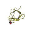
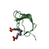
| Entry | Database: PDB / ID: 6b29 | ||||||
|---|---|---|---|---|---|---|---|
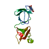
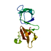
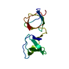
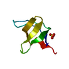
| Title | Crystal structure of the second SH3 domain of STAC3 (309-364) | ||||||
 Components Components | SH3 and cysteine-rich domain-containing protein 3 | ||||||
 Keywords Keywords |  PROTEIN BINDING / excitation-contraction coupling / ion channel adaptor protein PROTEIN BINDING / excitation-contraction coupling / ion channel adaptor protein | ||||||
| Function / homology |  Function and homology information Function and homology informationpositive regulation of voltage-gated calcium channel activity / neuromuscular synaptic transmission / voltage-gated calcium channel complex / skeletal muscle contraction / skeletal muscle fiber development / T-tubule / extrinsic component of cytoplasmic side of plasma membrane / positive regulation of protein localization to plasma membrane / cytoplasmic side of plasma membrane / synapse ...positive regulation of voltage-gated calcium channel activity / neuromuscular synaptic transmission / voltage-gated calcium channel complex / skeletal muscle contraction / skeletal muscle fiber development / T-tubule / extrinsic component of cytoplasmic side of plasma membrane / positive regulation of protein localization to plasma membrane / cytoplasmic side of plasma membrane / synapse / nucleoplasm / identical protein binding / metal ion binding / cytosolSimilarity search - Function | ||||||
| Biological species |  Homo sapiens (human) Homo sapiens (human) | ||||||
| Method | X-RAY DIFFRACTION / SYNCHROTRON / MOLECULAR REPLACEMENT / Resolution: 1.3 Å | ||||||
 Authors Authors | Wong King Yuen, S.M. / Van Petegem, F. | ||||||
| Funding support |  Canada, 1items Canada, 1items
| ||||||
 Citation Citation | Journal: Proc. Natl. Acad. Sci. U.S.A. / Year: 2017 Title: Structural insights into binding of STAC proteins to voltage-gated calcium channels. Authors: Wong King Yuen, S.M. / Campiglio, M. / Tung, C.C. / Flucher, B.E. / Van Petegem, F. | ||||||
| History |
|
- Structure visualization
| Structure viewer | Molecule: MolmilJmol/JSmol |
|---|
- Downloads & links
-Download
| PDBx/mmCIF format | 6b29.cif.gz | 109.2 KB | Display | PDBx/mmCIF format |
|---|---|---|---|---|
| PDB format | pdb6b29.ent.gz | 84.7 KB | Display | PDB format |
| PDBx/mmJSON format | 6b29.json.gz | Tree view | PDBx/mmJSON format | |
| Others |  Other downloads Other downloads |
-Validation report
| Arichive directory | https://data.pdbj.org/pub/pdb/validation_reports/b2/6b29ftp://data.pdbj.org/pub/pdb/validation_reports/b2/6b29 | HTTPS FTP |
|---|
-Related structure data


-Links
 PDBj
PDBj


- Assembly
| Deposited unit | 
| ||||||||
|---|---|---|---|---|---|---|---|---|---|
| 1 | 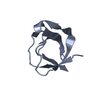
| ||||||||
| 2 | 
| ||||||||
| 3 | 
| ||||||||
| 4 | 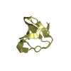
| ||||||||
| Unit cell |
|
-Components
| #1: Protein | Mass: 6632.563 Da / Num. of mol.: 4 / Fragment: residues 309-364 Source method: isolated from a genetically manipulated source Source: (gene. exp.) Homo sapiens (human) / Gene: STAC3 / Production host:  Escherichia coli (E. coli) / References: UniProt: Q96MF2 Escherichia coli (E. coli) / References: UniProt: Q96MF2#2: Water | ChemComp-HOH / | Water Mass: 18.015 Da / Num. of mol.: 305 / Source method: isolated from a natural source / Formula: H2O Mass: 18.015 Da / Num. of mol.: 305 / Source method: isolated from a natural source / Formula: H2O |
|---|
-Experimental details
-Experiment
| Experiment | Method: X-RAY DIFFRACTION / Number of used crystals: 1 |
|---|
- Sample preparation
| Crystal | Density Matthews: 2.12 Å3/Da / Density % sol: 42.02 % |
|---|---|
| Crystal grow | Temperature: 298 K / Method: vapor diffusion / pH: 6.5 Details: 0.1M Bis-Tris, 22.5% PEG3350 and 0.2M ammonium acetate |
-Data collection
| Diffraction | Mean temperature: 100 K |
|---|---|
| Diffraction source | Source: SYNCHROTRON / Site: CLSI / Beamline: 08ID-1 / Wavelength: 0.97949 Å |
| Detector | Type: DECTRIS PILATUS3 S 6M / Detector: PIXEL / Date: Aug 26, 2015 |
| Radiation | Protocol: SINGLE WAVELENGTH / Monochromatic (M) / Laue (L): M / Scattering type: x-ray |
| Radiation wavelength | Wavelength: 0.97949 Å / Relative weight: 1 |
| Reflection | Resolution: 1.3→39.205 Å / Num. obs: 55375 / % possible obs: 98.97 % / Redundancy: 6.7 % / Biso Wilson estimate: 14.57 Å2 / Rmerge(I) obs: 0.05199 / Net I/σ(I): 20.96 |
| Reflection shell | Resolution: 1.3→1.35 Å / Redundancy: 4.3 % / Rmerge(I) obs: 0.371 / Mean I/σ(I) obs: 3.41 / % possible all: 91.63 |
- Processing
| Software |
| |||||||||||||||||||||||||||||||||||||||||||||||||||||||||||||||||||||||||||||||||||||||||||||||||||||||||||||||||||||||||||||||||||||||||||||||||||
|---|---|---|---|---|---|---|---|---|---|---|---|---|---|---|---|---|---|---|---|---|---|---|---|---|---|---|---|---|---|---|---|---|---|---|---|---|---|---|---|---|---|---|---|---|---|---|---|---|---|---|---|---|---|---|---|---|---|---|---|---|---|---|---|---|---|---|---|---|---|---|---|---|---|---|---|---|---|---|---|---|---|---|---|---|---|---|---|---|---|---|---|---|---|---|---|---|---|---|---|---|---|---|---|---|---|---|---|---|---|---|---|---|---|---|---|---|---|---|---|---|---|---|---|---|---|---|---|---|---|---|---|---|---|---|---|---|---|---|---|---|---|---|---|---|---|---|---|---|
| Refinement | Method to determine structure: MOLECULAR REPLACEMENT / Resolution: 1.3→39.205 Å / SU ML: 0.12 / Cross valid method: FREE R-VALUE / σ(F): 1.37 / Phase error: 17.55
| |||||||||||||||||||||||||||||||||||||||||||||||||||||||||||||||||||||||||||||||||||||||||||||||||||||||||||||||||||||||||||||||||||||||||||||||||||
| Solvent computation | Shrinkage radii: 0.9 Å / VDW probe radii: 1.11 Å | |||||||||||||||||||||||||||||||||||||||||||||||||||||||||||||||||||||||||||||||||||||||||||||||||||||||||||||||||||||||||||||||||||||||||||||||||||
| Displacement parameters | Biso max: 86.9 Å2 / Biso mean: 21.7583 Å2 / Biso min: 8.09 Å2 | |||||||||||||||||||||||||||||||||||||||||||||||||||||||||||||||||||||||||||||||||||||||||||||||||||||||||||||||||||||||||||||||||||||||||||||||||||
| Refinement step | Cycle: final / Resolution: 1.3→39.205 Å
| |||||||||||||||||||||||||||||||||||||||||||||||||||||||||||||||||||||||||||||||||||||||||||||||||||||||||||||||||||||||||||||||||||||||||||||||||||
| Refine LS restraints |
| |||||||||||||||||||||||||||||||||||||||||||||||||||||||||||||||||||||||||||||||||||||||||||||||||||||||||||||||||||||||||||||||||||||||||||||||||||
| LS refinement shell | Refine-ID: X-RAY DIFFRACTION / Rfactor Rfree error: 0 / Total num. of bins used: 20
|
