- PDB-6b26: Crystal structure of human STAC2 Tandem SH3 Domains (296-411) -
+
Open data
ID or keywords:
Loading...
-
Basic information
Entry
Database: PDB / ID: 6b26
Title
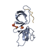
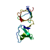
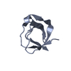
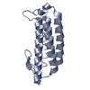
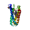
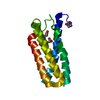
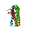
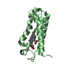
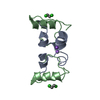
Crystal structure of human STAC2 Tandem SH3 Domains (296-411)
Components
SH3 and cysteine-rich domain-containing protein 2
Keywords
PROTEIN BINDING / Excitation-contraction coupling / ion channel adaptor protein
Function / homology
Function and homology information
positive regulation of voltage-gated calcium channel activity / skeletal muscle contraction / extrinsic component of cytoplasmic side of plasma membrane / positive regulation of protein localization to plasma membrane / sarcolemma / cytoplasmic side of plasma membrane / metal ion binding / cytosol Similarity search - Function
Stac2, SH3 domain / STAC1/2/3 / Unstructured on SH3 and cysteine-rich domain-containing protein 2 / Variant SH3 domain / Variant SH3 domain / Phorbol esters/diacylglycerol binding domain (C1 domain) / Zinc finger phorbol-ester/DAG-type signature. / Zinc finger phorbol-ester/DAG-type profile. / Protein kinase C conserved region 1 (C1) domains (Cysteine-rich domains) / Protein kinase C-like, phorbol ester/diacylglycerol-binding domain ...Stac2, SH3 domain / STAC1/2/3 / Unstructured on SH3 and cysteine-rich domain-containing protein 2 / Variant SH3 domain / Variant SH3 domain / Phorbol esters/diacylglycerol binding domain (C1 domain) / Zinc finger phorbol-ester/DAG-type signature. / Zinc finger phorbol-ester/DAG-type profile. / Protein kinase C conserved region 1 (C1) domains (Cysteine-rich domains) / Protein kinase C-like, phorbol ester/diacylglycerol-binding domain / C1-like domain superfamily / SH3 Domains / SH3 type barrels. / Src homology 3 domains / SH3-like domain superfamily / Src homology 3 (SH3) domain profile. / SH3 domain / Roll / Mainly Beta Similarity search - Domain/homology
Resolution: 1.2→32.95 Å / Cor.coef. Fo:Fc: 0.974 / Cor.coef. Fo:Fc free: 0.968 / SU B: 1.042 / SU ML: 0.022 / SU R Cruickshank DPI: 0.0393 / Cross valid method: THROUGHOUT / σ(F): 0 / ESU R: 0.039 / ESU R Free: 0.037 Details: HYDROGENS HAVE BEEN ADDED IN THE RIDING POSITIONS U VALUES : REFINED INDIVIDUALLY
Rfactor
Num. reflection
% reflection
Selection details
Rfree
0.1691
2040
4.9 %
RANDOM
Rwork
0.1524
-
-
-
obs
0.1532
39292
99.32 %
-
Solvent computation
Ion probe radii: 0.8 Å / Shrinkage radii: 0.8 Å / VDW probe radii: 1.2 Å
In the structure databanks used in Yorodumi, some data are registered as the other names, "COVID-19 virus" and "2019-nCoV". Here are the details of the virus and the list of structure data.
Jan 31, 2019. EMDB accession codes are about to change! (news from PDBe EMDB page)
EMDB accession codes are about to change! (news from PDBe EMDB page)
The allocation of 4 digits for EMDB accession codes will soon come to an end. Whilst these codes will remain in use, new EMDB accession codes will include an additional digit and will expand incrementally as the available range of codes is exhausted. The current 4-digit format prefixed with “EMD-” (i.e. EMD-XXXX) will advance to a 5-digit format (i.e. EMD-XXXXX), and so on. It is currently estimated that the 4-digit codes will be depleted around Spring 2019, at which point the 5-digit format will come into force.
The EM Navigator/Yorodumi systems omit the EMD- prefix.
Related info.:Q: What is EMD? / ID/Accession-code notation in Yorodumi/EM Navigator
Yorodumi is a browser for structure data from EMDB, PDB, SASBDB, etc.
This page is also the successor to EM Navigator detail page, and also detail information page/front-end page for Omokage search.
The word "yorodu" (or yorozu) is an old Japanese word meaning "ten thousand". "mi" (miru) is to see.
Related info.:EMDB / PDB / SASBDB / Comparison of 3 databanks / Yorodumi Search / Aug 31, 2016. New EM Navigator & Yorodumi / Yorodumi Papers / Jmol/JSmol / Function and homology information / Changes in new EM Navigator and Yorodumi
 Movie
Movie Controller


 Open data
 Basic information
 Components
 Keywords
 PROTEIN BINDING /
  Function and homology information
 Authors
 Canada, 1items
  Citation
 Structure visualization
 Downloads & links
 Other downloads
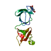


 PDBj
PDBj


 Assembly
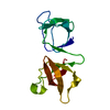

 Mass: 18.015 Da / Num. of mol.: 240 / Source method: isolated from a natural source / Formula: H2O
 Sample preparation
 / Beamline: BL9-2 / Wavelength: 0.97946 Å
 Processing