 Movie
Movie Controller

[English] 日本語
 Yorodumi
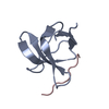
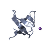
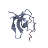
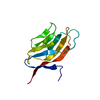
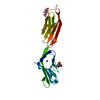
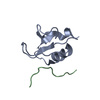
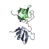
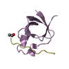
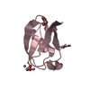
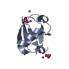
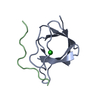
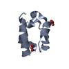
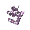
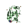
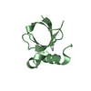
- PDB-2j7i: ATYPICAL POLYPROLINE RECOGNITION BY THE CMS N-TERMINAL SH3 DOMAIN... -
+ Open data

- Basic information
| Entry | Database: PDB / ID: 2j7i | ||||||
|---|---|---|---|---|---|---|---|
| Title | ATYPICAL POLYPROLINE RECOGNITION BY THE CMS N-TERMINAL SH3 DOMAIN. CMS:CD2 HETERODIMER | ||||||
 Components Components |
| ||||||
 Keywords Keywords |  PROTEIN BINDING / COILED COIL / POLYMORPHISM / GLYCOPROTEIN / CELL ADHESION / EGFR DOWNREGULATION / IMMUNOGLOBULIN DOMAIN / TRANSMEMBRANE / PHOSPHORYLATION / ADAPTOR PROTEIN / CMS / CD2AD / MEMBRANE / SH3 DOMAIN / SH3-BINDING / SH3 DOMAIN RECOGNITION PROTEIN BINDING / COILED COIL / POLYMORPHISM / GLYCOPROTEIN / CELL ADHESION / EGFR DOWNREGULATION / IMMUNOGLOBULIN DOMAIN / TRANSMEMBRANE / PHOSPHORYLATION / ADAPTOR PROTEIN / CMS / CD2AD / MEMBRANE / SH3 DOMAIN / SH3-BINDING / SH3 DOMAIN RECOGNITION | ||||||
| Function / homology |  Function and homology information Function and homology informationpositive regulation of myeloid dendritic cell activation / membrane raft polarization / negative regulation of small GTPase mediated signal transduction / transforming growth factor beta1 production / negative regulation of transforming growth factor beta1 production / response to transforming growth factor beta / immunological synapse formation / substrate-dependent cell migration, cell extension / protein heterooligomerization / natural killer cell mediated cytotoxicity ...positive regulation of myeloid dendritic cell activation / membrane raft polarization / negative regulation of small GTPase mediated signal transduction / transforming growth factor beta1 production / negative regulation of transforming growth factor beta1 production / response to transforming growth factor beta / immunological synapse formation / substrate-dependent cell migration, cell extension / protein heterooligomerization / natural killer cell mediated cytotoxicity / natural killer cell activation / podosome / Nephrin family interactions / heterotypic cell-cell adhesion / regulation of T cell differentiation / cell leading edge / filamentous actin / centriolar satellite / stress-activated MAPK cascade / ruffle / T cell activation / phosphatidylinositol 3-kinase/protein kinase B signal transduction / kidney development / actin filament organization / positive regulation of interleukin-8 production / positive regulation of protein secretion / regulation of actin cytoskeleton organization / Cell surface interactions at the vascular wall / synapse organization / protein catabolic process / neuromuscular junction / cytoplasmic side of plasma membrane / structural constituent of cytoskeleton / cell-cell adhesion / receptor tyrosine kinase binding / fibrillar center / SH3 domain binding / positive regulation of protein localization to nucleus / male gonad development / : / cell migration / actin filament binding / cell-cell junction / positive regulation of type II interferon production / actin cytoskeleton / late endosome / positive regulation of tumor necrosis factor production / signaling receptor activity / T cell receptor signaling pathway / protein-containing complex assembly / vesicle / cell surface receptor signaling pathway / cadherin binding / inflammatory response / cell cycle / axon / cell division / external side of plasma membrane / signaling receptor binding / apoptotic process / dendrite / Golgi apparatus / cell surface / signal transduction / protein-containing complex / extracellular exosome / extracellular region / nucleoplasm / identical protein binding / plasma membrane / cytoplasmSimilarity search - Function | ||||||
| Biological species |  HOMO SAPIENS (human) HOMO SAPIENS (human) | ||||||
| Method | X-RAY DIFFRACTION / MOLECULAR REPLACEMENT / Resolution: 2.9 Å | ||||||
 Authors Authors | Moncalian, G. / Cardenes, N. / Deribe, Y.L. / Spinola-Amilibia, M. / Dikic, I. / Bravo, J. | ||||||
 Citation Citation | Journal: J.Biol.Chem. / Year: 2006 Title: Atypical Polyproline Recognition by the Cms N-Terminal Src Homology 3 Domain. Authors: Moncalian, G. / Cardenes, N. / Deribe, Y.L. / Spinola-Amilibia, M. / Dikic, I. / Bravo, J. | ||||||
| History |
|
- Structure visualization
| Structure viewer | Molecule: MolmilJmol/JSmol |
|---|
- Downloads & links
-Download
| PDBx/mmCIF format | 2j7i.cif.gz | 39.8 KB | Display | PDBx/mmCIF format |
|---|---|---|---|---|
| PDB format | pdb2j7i.ent.gz | 28.7 KB | Display | PDB format |
| PDBx/mmJSON format | 2j7i.json.gz | Tree view | PDBx/mmJSON format | |
| Others |  Other downloads Other downloads |
-Validation report
| Arichive directory | https://data.pdbj.org/pub/pdb/validation_reports/j7/2j7iftp://data.pdbj.org/pub/pdb/validation_reports/j7/2j7i | HTTPS FTP |
|---|
-Related structure data


-Links
 PDBj
PDBj


- Assembly
| Deposited unit | 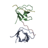
| ||||||||
|---|---|---|---|---|---|---|---|---|---|
| 1 | 
| ||||||||
| 2 | 
| ||||||||
| Unit cell |
|
-Components
| #1: Protein | Mass: 7412.285 Da / Num. of mol.: 2 / Fragment: SH3 DOMAIN, RESIDUES 1-62 Source method: isolated from a genetically manipulated source Details: N-TERMINAL SH3 DOMAIN FROM CMS (CAS LIGAND WITH MULTIPLE SH3 DOMAINS) OR CD2AP (CD2-ASSOCIATED PROTEIN) Source: (gene. exp.) HOMO SAPIENS (human) / Plasmid: PET 21A / Production host:  ESCHERICHIA COLI (E. coli) / Strain (production host): ROSSETTA (DE3) PLYS / References: UniProt: Q9Y5K6 ESCHERICHIA COLI (E. coli) / Strain (production host): ROSSETTA (DE3) PLYS / References: UniProt: Q9Y5K6#2: Protein/peptide | Mass: 1119.382 Da / Num. of mol.: 2 / Fragment: CMS BINDING SEQUENCE, RESIDUES 324-333 / Source method: obtained synthetically / Source: (synth.) HOMO SAPIENS (human) / References: UniProt: P06729#3: Water | ChemComp-HOH / | Water Mass: 18.015 Da / Num. of mol.: 31 / Source method: isolated from a natural source / Formula: H2O Mass: 18.015 Da / Num. of mol.: 31 / Source method: isolated from a natural source / Formula: H2O |
|---|
-Experimental details
-Experiment
| Experiment | Method: X-RAY DIFFRACTION / Number of used crystals: 1 |
|---|
- Sample preparation
| Crystal | Density Matthews: 2.6 Å3/Da / Density % sol: 52.2 % |
|---|---|
| Crystal grow | pH: 8 / Details: 30% PEG8000, 0.2M NACL, 0.1M TRIS-HCL PH 8.0 |
-Data collection
| Diffraction | Mean temperature: 110 K |
|---|---|
| Diffraction source | Source: ROTATING ANODE / Type: ENRAF-NONIUS FR591 / Wavelength: 1.54179 |
| Detector | Type: MARRESERACH / Detector: IMAGE PLATE / Date: Jun 8, 2004 / Details: MIRRORS |
| Radiation | Protocol: SINGLE WAVELENGTH / Monochromatic (M) / Laue (L): M / Scattering type: x-ray |
| Radiation wavelength | Wavelength: 1.54179 Å / Relative weight: 1 |
| Reflection | Resolution: 2.9→36.29 Å / Num. obs: 3729 / % possible obs: 100 % / Observed criterion σ(I): 2.4 / Redundancy: 8 % / Biso Wilson estimate: 49.9 Å2 / Rmerge(I) obs: 0.1 / Net I/σ(I): 22 |
| Reflection shell | Resolution: 2.9→3.1 Å / Redundancy: 8.1 % / Rmerge(I) obs: 0.31 / Mean I/σ(I) obs: 6.2 / % possible all: 100 |
- Processing
| Software |
| ||||||||||||||||||||||||||||||||||||||||||||||||||||||||||||||||||||||||||||||||
|---|---|---|---|---|---|---|---|---|---|---|---|---|---|---|---|---|---|---|---|---|---|---|---|---|---|---|---|---|---|---|---|---|---|---|---|---|---|---|---|---|---|---|---|---|---|---|---|---|---|---|---|---|---|---|---|---|---|---|---|---|---|---|---|---|---|---|---|---|---|---|---|---|---|---|---|---|---|---|---|---|---|
| Refinement | Method to determine structure: MOLECULAR REPLACEMENT Starting model: CMSA-CBL-B STRUCTURE Resolution: 2.9→19.8 Å / Rfactor Rfree error: 0.022 / Data cutoff high absF: 590838.33 / Isotropic thermal model: RESTRAINED / Cross valid method: THROUGHOUT / σ(F): 0
| ||||||||||||||||||||||||||||||||||||||||||||||||||||||||||||||||||||||||||||||||
| Solvent computation | Solvent model: FLAT MODEL / Bsol: 24.9924 Å2 / ksol: 0.320003 e/Å3 | ||||||||||||||||||||||||||||||||||||||||||||||||||||||||||||||||||||||||||||||||
| Displacement parameters | Biso mean: 38.2 Å2
| ||||||||||||||||||||||||||||||||||||||||||||||||||||||||||||||||||||||||||||||||
| Refine analyze |
| ||||||||||||||||||||||||||||||||||||||||||||||||||||||||||||||||||||||||||||||||
| Refinement step | Cycle: LAST / Resolution: 2.9→19.8 Å
| ||||||||||||||||||||||||||||||||||||||||||||||||||||||||||||||||||||||||||||||||
| Refine LS restraints |
| ||||||||||||||||||||||||||||||||||||||||||||||||||||||||||||||||||||||||||||||||
| LS refinement shell | Resolution: 2.9→3.08 Å / Rfactor Rfree error: 0.076 / Total num. of bins used: 6
| ||||||||||||||||||||||||||||||||||||||||||||||||||||||||||||||||||||||||||||||||
| Xplor file |
|
