 Movie
Movie Controller


+ Open data

- Basic information
| Entry | Database: PDB / ID: 2f7d | ||||||
|---|---|---|---|---|---|---|---|
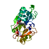
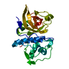
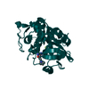
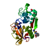
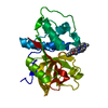
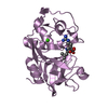
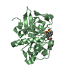
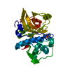
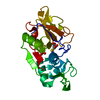
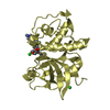
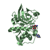
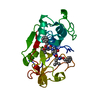
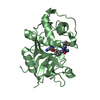
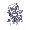
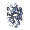
| Title | A mutant rabbit cathepsin K with a nitrile inhibitor | ||||||
 Components Components | Cathepsin K | ||||||
 Keywords Keywords | HYDROLASE / papain cysteine protease | ||||||
| Function / homology |  Function and homology informationcathepsin K / negative regulation of cartilage development / thyroid hormone generation / collagen catabolic process / bone resorption / proteolysis involved in protein catabolic process / lysosome / apical plasma membrane / external side of plasma membrane / cysteine-type endopeptidase activity / extracellular space Function and homology informationcathepsin K / negative regulation of cartilage development / thyroid hormone generation / collagen catabolic process / bone resorption / proteolysis involved in protein catabolic process / lysosome / apical plasma membrane / external side of plasma membrane / cysteine-type endopeptidase activity / extracellular spaceSimilarity search - Function | ||||||
| Biological species |  Oryctolagus cuniculus (rabbit) Oryctolagus cuniculus (rabbit) | ||||||
| Method | X-RAY DIFFRACTION / FOURIER SYNTHESIS / Resolution: 1.9 Å | ||||||
 Authors Authors | Somoza, J.R. | ||||||
 Citation Citation | Journal: J.Med.Chem. / Year: 2006 Title: Beta-substituted cyclohexanecarboxamide: a nonpeptidic framework for the design of potent inhibitors of cathepsin K. Authors: Crane, S.N. / Black, W.C. / Palmer, J.T. / Davis, D.E. / Setti, E. / Robichaud, J. / Paquet, J. / Oballa, R.M. / Bayly, C.I. / McKay, D.J. / Somoza, J.R. / Chauret, N. / Seto, C. / ...Authors: Crane, S.N. / Black, W.C. / Palmer, J.T. / Davis, D.E. / Setti, E. / Robichaud, J. / Paquet, J. / Oballa, R.M. / Bayly, C.I. / McKay, D.J. / Somoza, J.R. / Chauret, N. / Seto, C. / Scheigetz, J. / Wesolowski, G. / Masse, F. / Desmarais, S. / Ouellet, M. | ||||||
| History |
|
- Structure visualization
| Structure viewer | Molecule: MolmilJmol/JSmol |
|---|
- Downloads & links
-Download
| PDBx/mmCIF format | 2f7d.cif.gz | 53.7 KB | Display | PDBx/mmCIF format |
|---|---|---|---|---|
| PDB format | pdb2f7d.ent.gz | 40.5 KB | Display | PDB format |
| PDBx/mmJSON format | 2f7d.json.gz | Tree view | PDBx/mmJSON format | |
| Others |  Other downloads Other downloads |
-Validation report
| Arichive directory | https://data.pdbj.org/pub/pdb/validation_reports/f7/2f7dftp://data.pdbj.org/pub/pdb/validation_reports/f7/2f7d | HTTPS FTP |
|---|
-Related structure data
| Similar structure data |
|---|

-Links
 PDBj
PDBj
- Assembly
| Deposited unit | 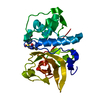
| ||||||||
|---|---|---|---|---|---|---|---|---|---|
| 1 |
| ||||||||
| Unit cell |
| ||||||||
| Details | The cathepsin K monomer is probably the biological unit. |
-Components
| #1: Protein | / OC-2 protein Mass: 23554.498 Da / Num. of mol.: 1 / Mutation: Y61D, V157L Source method: isolated from a genetically manipulated source Source: (gene. exp.) Oryctolagus cuniculus (rabbit) / Gene: CTSK / Production host:  Pichia pastoris (fungus) / References: UniProt: P43236, cathepsin K Pichia pastoris (fungus) / References: UniProt: P43236, cathepsin K |
|---|---|
| #2: Chemical | ChemComp-NOQ / (  Mass: 354.464 Da / Num. of mol.: 1 / Source method: obtained synthetically / Formula: C17H26N2O4S Mass: 354.464 Da / Num. of mol.: 1 / Source method: obtained synthetically / Formula: C17H26N2O4S |
| #3: Water | ChemComp-HOH / Water Mass: 18.015 Da / Num. of mol.: 97 / Source method: isolated from a natural source / Formula: H2O Mass: 18.015 Da / Num. of mol.: 97 / Source method: isolated from a natural source / Formula: H2O |
-Experimental details
-Experiment
| Experiment | Method: X-RAY DIFFRACTION / Number of used crystals: 1 |
|---|
- Sample preparation
| Crystal | Density Matthews: 2.16 Å3/Da / Density % sol: 42.97 % |
|---|---|
| Crystal grow | Temperature: 291 K / Method: vapor diffusion, hanging drop / pH: 3.9 Details: 0.18 M magnesium formate, pH 3.9, VAPOR DIFFUSION, HANGING DROP, temperature 291K |
-Data collection
| Diffraction | Mean temperature: 298 K | ||||||||||||||||||||||||||||||||||||||||||||||||||||||||||||||||||
|---|---|---|---|---|---|---|---|---|---|---|---|---|---|---|---|---|---|---|---|---|---|---|---|---|---|---|---|---|---|---|---|---|---|---|---|---|---|---|---|---|---|---|---|---|---|---|---|---|---|---|---|---|---|---|---|---|---|---|---|---|---|---|---|---|---|---|---|
| Diffraction source | Source: ROTATING ANODE / Type: RIGAKU RU200 / Wavelength: 1.54 Å | ||||||||||||||||||||||||||||||||||||||||||||||||||||||||||||||||||
| Detector | Type: RIGAKU RAXIS IV / Detector: IMAGE PLATE / Date: Sep 11, 2001 | ||||||||||||||||||||||||||||||||||||||||||||||||||||||||||||||||||
| Radiation | Protocol: SINGLE WAVELENGTH / Monochromatic (M) / Laue (L): M / Scattering type: x-ray | ||||||||||||||||||||||||||||||||||||||||||||||||||||||||||||||||||
| Radiation wavelength | Wavelength: 1.54 Å / Relative weight: 1 | ||||||||||||||||||||||||||||||||||||||||||||||||||||||||||||||||||
| Reflection | Number: 16445 / Rmerge(I) obs: 0.081 / Χ2: 1.133 / D res high: 1.9 Å / D res low: 100 Å / % possible obs: 98.1 | ||||||||||||||||||||||||||||||||||||||||||||||||||||||||||||||||||
| Diffraction reflection shell |
| ||||||||||||||||||||||||||||||||||||||||||||||||||||||||||||||||||
| Reflection | Resolution: 1.9→100 Å / Num. all: 16445 / Num. obs: 16445 / % possible obs: 98.1 % / Observed criterion σ(F): 0 / Observed criterion σ(I): -3 / Rmerge(I) obs: 0.081 / Χ2: 1.133 | ||||||||||||||||||||||||||||||||||||||||||||||||||||||||||||||||||
| Reflection shell |
|
- Processing
| Software |
| |||||||||||||||||||||||||
|---|---|---|---|---|---|---|---|---|---|---|---|---|---|---|---|---|---|---|---|---|---|---|---|---|---|---|
| Refinement | Method to determine structure: FOURIER SYNTHESIS / Resolution: 1.9→36.51 Å / σ(F): 0 / Stereochemistry target values: Engh & Huber
| |||||||||||||||||||||||||
| Displacement parameters | Biso mean: 22.12 Å2
| |||||||||||||||||||||||||
| Refine analyze |
| |||||||||||||||||||||||||
| Refinement step | Cycle: LAST / Resolution: 1.9→36.51 Å
| |||||||||||||||||||||||||
| Refine LS restraints |
| |||||||||||||||||||||||||
| LS refinement shell | Resolution: 1.9→2.02 Å / Rfactor Rfree error: 0.016
| |||||||||||||||||||||||||
| Xplor file |
|
