 Movie
Movie Controller

[English] 日本語
 Yorodumi
- PDB-2ftd: Crystal structure of Cathepsin K complexed with 7-Methyl-Substitu... -
+ Open data

- Basic information
| Entry | Database: PDB / ID: 2ftd | ||||||
|---|---|---|---|---|---|---|---|
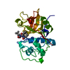
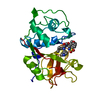
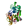
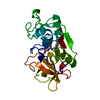
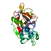
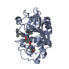
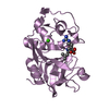
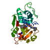
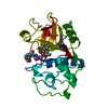
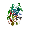
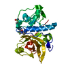
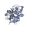
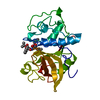
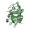
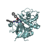
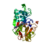
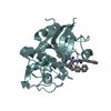
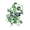
| Title | Crystal structure of Cathepsin K complexed with 7-Methyl-Substituted Azepan-3-one compound | ||||||
 Components Components | Cathepsin K | ||||||
 Keywords Keywords | HYDROLASE / Sulfhydryl Proteinase | ||||||
| Function / homology |  Function and homology informationcathepsin K / thyroid hormone generation / proteolysis involved in protein catabolic process / lysosome / immune response / apical plasma membrane / cysteine-type endopeptidase activity / extracellular space Function and homology informationcathepsin K / thyroid hormone generation / proteolysis involved in protein catabolic process / lysosome / immune response / apical plasma membrane / cysteine-type endopeptidase activity / extracellular spaceSimilarity search - Function | ||||||
| Biological species |  Macaca mulatta (Rhesus monkey) Macaca mulatta (Rhesus monkey) | ||||||
| Method | X-RAY DIFFRACTION / SYNCHROTRON / FOURIER SYNTHESIS / Resolution: 2.55 Å | ||||||
 Authors Authors | Yamashita, D.S. / Baoguang, Z. | ||||||
 Citation Citation | Journal: J.Med.Chem. / Year: 2006 Title: Structure activity relationships of 5-, 6-, and 7-methyl-substituted azepan-3-one cathepsin K inhibitors. Authors: Yamashita, D.S. / Marquis, R.W. / Xie, R. / Nidamarthy, S.D. / Oh, H.J. / Jeong, J.U. / Erhard, K.F. / Ward, K.W. / Roethke, T.J. / Smith, B.R. / Cheng, H.Y. / Geng, X. / Lin, F. / Offen, P. ...Authors: Yamashita, D.S. / Marquis, R.W. / Xie, R. / Nidamarthy, S.D. / Oh, H.J. / Jeong, J.U. / Erhard, K.F. / Ward, K.W. / Roethke, T.J. / Smith, B.R. / Cheng, H.Y. / Geng, X. / Lin, F. / Offen, P.H. / Wang, B. / Nevins, N. / Head, M.S. / Haltiwanger, R.C. / Narducci Sarjeant, A.A. / Liable-Sands, L.M. / Zhao, B. / Smith, W.W. / Janson, C.A. / Gao, E. / Tomaszek, T. / McQueney, M. / James, I.E. / Gress, C.J. / Zembryki, D.L. / Lark, M.W. / Veber, D.F. | ||||||
| History |
|
- Structure visualization
| Structure viewer | Molecule: MolmilJmol/JSmol |
|---|
- Downloads & links
-Download
| PDBx/mmCIF format | 2ftd.cif.gz | 94.4 KB | Display | PDBx/mmCIF format |
|---|---|---|---|---|
| PDB format | pdb2ftd.ent.gz | 78 KB | Display | PDB format |
| PDBx/mmJSON format | 2ftd.json.gz | Tree view | PDBx/mmJSON format | |
| Others |  Other downloads Other downloads |
-Validation report
| Arichive directory | https://data.pdbj.org/pub/pdb/validation_reports/ft/2ftdftp://data.pdbj.org/pub/pdb/validation_reports/ft/2ftd | HTTPS FTP |
|---|
-Related structure data
| Related structure data | |
|---|---|
| Similar structure data |


-Links
 PDBj
PDBj
- Assembly
| Deposited unit | 
| ||||||||
|---|---|---|---|---|---|---|---|---|---|
| 1 | 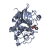
| ||||||||
| 2 | 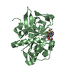
| ||||||||
| Unit cell |
|
-Components
| #1: Protein | Mass: 23523.480 Da / Num. of mol.: 2 Source method: isolated from a genetically manipulated source Source: (gene. exp.) Macaca mulatta (Rhesus monkey) / Strain: Osteoclast / Gene: CTSK / Production host:  unidentified baculovirus / Strain (production host): SF9 / References: UniProt: P61277, cathepsin K unidentified baculovirus / Strain (production host): SF9 / References: UniProt: P61277, cathepsin K#2: Chemical |   Mass: 540.631 Da / Num. of mol.: 2 / Source method: obtained synthetically / Formula: C27H32N4O6S Mass: 540.631 Da / Num. of mol.: 2 / Source method: obtained synthetically / Formula: C27H32N4O6S#3: Water | ChemComp-HOH / | Water Mass: 18.015 Da / Num. of mol.: 193 / Source method: isolated from a natural source / Formula: H2O Mass: 18.015 Da / Num. of mol.: 193 / Source method: isolated from a natural source / Formula: H2O |
|---|
-Experimental details
-Experiment
| Experiment | Method: X-RAY DIFFRACTION / Number of used crystals: 1 |
|---|
- Sample preparation
| Crystal | Density Matthews: 2.92 Å3/Da / Density % sol: 57.86 % |
|---|---|
| Crystal grow | Temperature: 283 K / Method: vapor diffusion, sitting drop / pH: 7.5 Details: 28% PEG 400 as precipitant, in 0.1 M Hepes buffer, at pH 7.5 containing 0.2 M CaCl2, temperature 283K, VAPOR DIFFUSION, SITTING DROP |
-Data collection
| Diffraction | Mean temperature: 93 K |
|---|---|
| Diffraction source | Source: SYNCHROTRON / Site: APS  / Beamline: 17-ID / Beamline: 17-ID |
| Detector | Detector: CCD / Date: Apr 1, 2002 |
| Radiation | Protocol: SINGLE WAVELENGTH / Monochromatic (M) / Laue (L): M / Scattering type: x-ray |
| Radiation wavelength | Relative weight: 1 |
| Reflection | Resolution: 2.55→100 Å / Num. all: 19480 / Num. obs: 17372 / % possible obs: 89.2 % / Observed criterion σ(F): 2 / Observed criterion σ(I): 2 / Redundancy: 9.9 % / Rsym value: 0.058 / Net I/σ(I): 19 |
| Reflection shell | Resolution: 2.55→2.59 Å / Mean I/σ(I) obs: 10.2 / Num. unique all: 631 / Rsym value: 0.094 / % possible all: 65 |
- Processing
| Software |
| ||||||||||||||||||||
|---|---|---|---|---|---|---|---|---|---|---|---|---|---|---|---|---|---|---|---|---|---|
| Refinement | Method to determine structure: FOURIER SYNTHESIS / Resolution: 2.55→25 Å / σ(F): 1 / Stereochemistry target values: Engh & Huber
| ||||||||||||||||||||
| Refinement step | Cycle: LAST / Resolution: 2.55→25 Å
| ||||||||||||||||||||
| Refine LS restraints |
|
