 Movie
Movie Controller

[English] 日本語
 Yorodumi
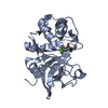
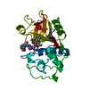
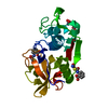
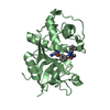
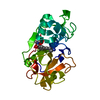
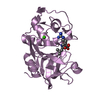
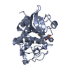
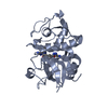
- PDB-3ovz: Cathepsin K in complex with a covalent inhibitor with a ketoamide... -
+ Open data

- Basic information
| Entry | Database: PDB / ID: 3ovz | ||||||
|---|---|---|---|---|---|---|---|
| Title | Cathepsin K in complex with a covalent inhibitor with a ketoamide warhead | ||||||
 Components Components | Cathepsin K | ||||||
 Keywords Keywords | HYDROLASE/HYDROLASE INHIBITOR / Cathepsin K / hydrolase / covalent inhibitor / ketoamide warhead / Ligand forms covalent bond to Cys25 / Lysosomes / HYDROLASE-HYDROLASE INHIBITOR complex | ||||||
| Function / homology |  Function and homology informationcathepsin K / mononuclear cell differentiation / intramembranous ossification / negative regulation of cartilage development / cellular response to zinc ion starvation / RUNX1 regulates transcription of genes involved in differentiation of keratinocytes / thyroid hormone generation / endolysosome lumen / Trafficking and processing of endosomal TLR / proteoglycan binding ...cathepsin K / mononuclear cell differentiation / intramembranous ossification / negative regulation of cartilage development / cellular response to zinc ion starvation / RUNX1 regulates transcription of genes involved in differentiation of keratinocytes / thyroid hormone generation / endolysosome lumen / Trafficking and processing of endosomal TLR / proteoglycan binding / Activation of Matrix Metalloproteinases / cysteine-type endopeptidase activator activity involved in apoptotic process / mitophagy / fibronectin binding / Collagen degradation / collagen catabolic process / extracellular matrix disassembly / bone resorption / cysteine-type peptidase activity / cellular response to transforming growth factor beta stimulus / collagen binding / MHC class II antigen presentation / Degradation of the extracellular matrix / lysosomal lumen / proteolysis involved in protein catabolic process / positive regulation of apoptotic signaling pathway / response to insulin / response to organic cyclic compound / cellular response to tumor necrosis factor / response to ethanol / lysosome / immune response / apical plasma membrane / external side of plasma membrane / cysteine-type endopeptidase activity / serine-type endopeptidase activity / intracellular membrane-bounded organelle / proteolysis / extracellular space / extracellular region / nucleoplasm Function and homology informationcathepsin K / mononuclear cell differentiation / intramembranous ossification / negative regulation of cartilage development / cellular response to zinc ion starvation / RUNX1 regulates transcription of genes involved in differentiation of keratinocytes / thyroid hormone generation / endolysosome lumen / Trafficking and processing of endosomal TLR / proteoglycan binding ...cathepsin K / mononuclear cell differentiation / intramembranous ossification / negative regulation of cartilage development / cellular response to zinc ion starvation / RUNX1 regulates transcription of genes involved in differentiation of keratinocytes / thyroid hormone generation / endolysosome lumen / Trafficking and processing of endosomal TLR / proteoglycan binding / Activation of Matrix Metalloproteinases / cysteine-type endopeptidase activator activity involved in apoptotic process / mitophagy / fibronectin binding / Collagen degradation / collagen catabolic process / extracellular matrix disassembly / bone resorption / cysteine-type peptidase activity / cellular response to transforming growth factor beta stimulus / collagen binding / MHC class II antigen presentation / Degradation of the extracellular matrix / lysosomal lumen / proteolysis involved in protein catabolic process / positive regulation of apoptotic signaling pathway / response to insulin / response to organic cyclic compound / cellular response to tumor necrosis factor / response to ethanol / lysosome / immune response / apical plasma membrane / external side of plasma membrane / cysteine-type endopeptidase activity / serine-type endopeptidase activity / intracellular membrane-bounded organelle / proteolysis / extracellular space / extracellular region / nucleoplasmSimilarity search - Function | ||||||
| Biological species |  Homo sapiens (human) Homo sapiens (human) | ||||||
| Method | X-RAY DIFFRACTION / MOLECULAR REPLACEMENT / Resolution: 2.02 Å | ||||||
 Authors Authors | Fradera, X. / van Zeeland, M. / Uitdehaag, J.C.M. | ||||||
 Citation Citation | Journal: Bioorg.Med.Chem.Lett. / Year: 2010 Title: Trifluoromethylphenyl as P2 for ketoamide-based cathepsin S inhibitors. Authors: Cai, J. / Robinson, J. / Belshaw, S. / Everett, K. / Fradera, X. / van Zeeland, M. / van Berkom, L. / van Rijnsbergen, P. / Popplestone, L. / Baugh, M. / Dempster, M. / Bruin, J. / Hamilton, ...Authors: Cai, J. / Robinson, J. / Belshaw, S. / Everett, K. / Fradera, X. / van Zeeland, M. / van Berkom, L. / van Rijnsbergen, P. / Popplestone, L. / Baugh, M. / Dempster, M. / Bruin, J. / Hamilton, W. / Kinghorn, E. / Westwood, P. / Kerr, J. / Rankovic, Z. / Arbuckle, W. / Bennett, D.J. / Jones, P.S. / Long, C. / Martin, I. / Uitdehaag, J.C. / Meulemans, T. | ||||||
| History |
|
- Structure visualization
| Structure viewer | Molecule: MolmilJmol/JSmol |
|---|
- Downloads & links
-Download
| PDBx/mmCIF format | 3ovz.cif.gz | 58.1 KB | Display | PDBx/mmCIF format |
|---|---|---|---|---|
| PDB format | pdb3ovz.ent.gz | 41.1 KB | Display | PDB format |
| PDBx/mmJSON format | 3ovz.json.gz | Tree view | PDBx/mmJSON format | |
| Others |  Other downloads Other downloads |
-Validation report
| Arichive directory | https://data.pdbj.org/pub/pdb/validation_reports/ov/3ovzftp://data.pdbj.org/pub/pdb/validation_reports/ov/3ovz | HTTPS FTP |
|---|
-Related structure data


-Links
 PDBj
PDBj


- Assembly
| Deposited unit | 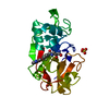
| ||||||||
|---|---|---|---|---|---|---|---|---|---|
| 1 |
| ||||||||
| Unit cell |
|
-Components
| #1: Protein | / Cathepsin O / Cathepsin O2 / Cathepsin X Mass: 23355.287 Da / Num. of mol.: 1 / Fragment: UNP residues 121-329 Source method: isolated from a genetically manipulated source Source: (gene. exp.) Homo sapiens (human) / Gene: CTSK, CTSO, CTSO2 / References: UniProt: P43235, cathepsin K | ||
|---|---|---|---|
| #2: Chemical | ChemComp-O96 /   Mass: 443.804 Da / Num. of mol.: 1 / Source method: obtained synthetically / Formula: C19H17ClF3N3O4 Mass: 443.804 Da / Num. of mol.: 1 / Source method: obtained synthetically / Formula: C19H17ClF3N3O4 | ||
| #3: Chemical | Sulfate  Mass: 96.063 Da / Num. of mol.: 2 / Source method: obtained synthetically / Formula: SO4 Mass: 96.063 Da / Num. of mol.: 2 / Source method: obtained synthetically / Formula: SO4#4: Water | ChemComp-HOH / | Water Mass: 18.015 Da / Num. of mol.: 79 / Source method: isolated from a natural source / Formula: H2O Mass: 18.015 Da / Num. of mol.: 79 / Source method: isolated from a natural source / Formula: H2O |
-Experimental details
-Experiment
| Experiment | Method: X-RAY DIFFRACTION / Number of used crystals: 1 |
|---|
- Sample preparation
| Crystal | Density Matthews: 2.02 Å3/Da / Density % sol: 38.61 % |
|---|---|
| Crystal grow | Temperature: 298 K Details: 0 mM NaAcetate pH=4.0, 0.3 M NaCl, 20% PEG4000, 0.2 M (NH4)2SO4, pH=2.9, 4% Methanol, Cryoprotectant composition:20% PEG4000, 0.1M (NH4)2SO4 pH=2.9, 4% Methanol, 20% PEG 400, pH 5, ...Details: 0 mM NaAcetate pH=4.0, 0.3 M NaCl, 20% PEG4000, 0.2 M (NH4)2SO4, pH=2.9, 4% Methanol, Cryoprotectant composition:20% PEG4000, 0.1M (NH4)2SO4 pH=2.9, 4% Methanol, 20% PEG 400, pH 5, cocrystallization, hanging drop, temperature 398K |
-Data collection
| Diffraction | Mean temperature: 100 K |
|---|---|
| Diffraction source | Source: ROTATING ANODE / Type: RIGAKU MICROMAX-007 HF / Wavelength: 1.5418 / Wavelength: 1.5418 Å |
| Detector | Type: RIGAKU RAXIS IV / Detector: IMAGE PLATE / Date: Feb 12, 2008 |
| Radiation | Protocol: SINGLE WAVELENGTH / Monochromatic (M) / Laue (L): M / Scattering type: x-ray |
| Radiation wavelength | Wavelength: 1.5418 Å / Relative weight: 1 |
| Reflection | Resolution: 2.02→55.74 Å / Num. obs: 14051 / % possible obs: 100 % |
| Reflection shell | Resolution: 2.02→2.09 Å / Rmerge(I) obs: 0.7 / Mean I/σ(I) obs: 2 / % possible all: 100 |
- Processing
| Software |
| ||||||||||||||||||||||||||||||||||||||||||||||||||||||||||||||||||||||||||||||||||||||||||||||||||||||||||||||||||||||||||||||||||||||||||||||||||||||||||||||||||||||||||
|---|---|---|---|---|---|---|---|---|---|---|---|---|---|---|---|---|---|---|---|---|---|---|---|---|---|---|---|---|---|---|---|---|---|---|---|---|---|---|---|---|---|---|---|---|---|---|---|---|---|---|---|---|---|---|---|---|---|---|---|---|---|---|---|---|---|---|---|---|---|---|---|---|---|---|---|---|---|---|---|---|---|---|---|---|---|---|---|---|---|---|---|---|---|---|---|---|---|---|---|---|---|---|---|---|---|---|---|---|---|---|---|---|---|---|---|---|---|---|---|---|---|---|---|---|---|---|---|---|---|---|---|---|---|---|---|---|---|---|---|---|---|---|---|---|---|---|---|---|---|---|---|---|---|---|---|---|---|---|---|---|---|---|---|---|---|---|---|---|---|---|---|
| Refinement | Method to determine structure: MOLECULAR REPLACEMENT Starting model: In-house cathepsin K structure Resolution: 2.02→51.16 Å / Cor.coef. Fo:Fc: 0.934 / Cor.coef. Fo:Fc free: 0.902 / SU B: 7.597 / SU ML: 0.204 / Stereochemistry target values: MAXIMUM LIKELIHOOD / Details: HYDROGENS HAVE BEEN ADDED IN THE RIDING POSITIONS
| ||||||||||||||||||||||||||||||||||||||||||||||||||||||||||||||||||||||||||||||||||||||||||||||||||||||||||||||||||||||||||||||||||||||||||||||||||||||||||||||||||||||||||
| Solvent computation | Solvent model: MASK | ||||||||||||||||||||||||||||||||||||||||||||||||||||||||||||||||||||||||||||||||||||||||||||||||||||||||||||||||||||||||||||||||||||||||||||||||||||||||||||||||||||||||||
| Displacement parameters | Biso mean: 35.26 Å2
| ||||||||||||||||||||||||||||||||||||||||||||||||||||||||||||||||||||||||||||||||||||||||||||||||||||||||||||||||||||||||||||||||||||||||||||||||||||||||||||||||||||||||||
| Refinement step | Cycle: LAST / Resolution: 2.02→51.16 Å
| ||||||||||||||||||||||||||||||||||||||||||||||||||||||||||||||||||||||||||||||||||||||||||||||||||||||||||||||||||||||||||||||||||||||||||||||||||||||||||||||||||||||||||
| Refine LS restraints |
| ||||||||||||||||||||||||||||||||||||||||||||||||||||||||||||||||||||||||||||||||||||||||||||||||||||||||||||||||||||||||||||||||||||||||||||||||||||||||||||||||||||||||||
| LS refinement shell | Resolution: 2.02→2.07 Å
|
