 Movie
Movie Controller


+ Open data

- Basic information
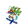
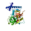
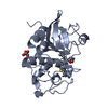
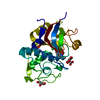
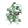
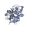
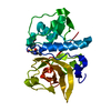
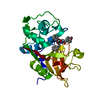
| Entry | Database: PDB / ID: 2xu1 | ||||||
|---|---|---|---|---|---|---|---|
| Title | CATHEPSIN L WITH A NITRILE INHIBITOR | ||||||
 Components Components | CATHEPSIN L1 | ||||||
 Keywords Keywords | HYDROLASE / DRUG DESIGN / THIOL PROTEASE | ||||||
| Function / homology |  Function and homology information Function and homology informationenkephalin processing / cathepsin L / CD4-positive, alpha-beta T cell lineage commitment / macrophage apoptotic process / chromaffin granule / elastin catabolic process / antigen processing and presentation of peptide antigen / RUNX1 regulates transcription of genes involved in differentiation of keratinocytes / endolysosome lumen / cellular response to thyroid hormone stimulus ...enkephalin processing / cathepsin L / CD4-positive, alpha-beta T cell lineage commitment / macrophage apoptotic process / chromaffin granule / elastin catabolic process / antigen processing and presentation of peptide antigen / RUNX1 regulates transcription of genes involved in differentiation of keratinocytes / endolysosome lumen / cellular response to thyroid hormone stimulus / zymogen activation / Trafficking and processing of endosomal TLR / proteoglycan binding / Assembly of collagen fibrils and other multimeric structures / cysteine-type endopeptidase activator activity involved in apoptotic process / protein autoprocessing / Collagen degradation / fibronectin binding / antigen processing and presentation / collagen catabolic process / serpin family protein binding / cysteine-type peptidase activity / Attachment and Entry / positive regulation of apoptotic signaling pathway / endocytic vesicle lumen / collagen binding / MHC class II antigen presentation / Degradation of the extracellular matrix / multivesicular body / proteolysis involved in protein catabolic process / lysosomal lumen / Endosomal/Vacuolar pathway / antigen processing and presentation of exogenous peptide antigen via MHC class II / histone binding / collagen-containing extracellular matrix / receptor-mediated endocytosis of virus by host cell / Attachment and Entry / adaptive immune response / lysosome / immune response / symbiont entry into host cell / apical plasma membrane / fusion of virus membrane with host plasma membrane / cysteine-type endopeptidase activity / intracellular membrane-bounded organelle / fusion of virus membrane with host endosome membrane / Golgi apparatus / proteolysis / extracellular space / extracellular exosome / extracellular region / nucleus / plasma membraneSimilarity search - Function | ||||||
| Biological species |  HOMO SAPIENS (human) HOMO SAPIENS (human) | ||||||
| Method | X-RAY DIFFRACTION / SYNCHROTRON / OTHER / Resolution: 1.45 Å | ||||||
 Authors Authors | Banner, D.W. / Benz, J.M. / Steinbacher, S. / Haap, W. | ||||||
 Citation Citation | Journal: Angew.Chem.Int.Ed.Engl. / Year: 2011 Title: Systematic Investigation of Halogen Bonding in Protein-Ligand Interactions. Authors: Hardegger, L.A. / Kuhn, B. / Spinnler, B. / Anselm, L. / Ecabert, R. / Stihle, M. / Gsell, B. / Thoma, R. / Diez, J. / Benz, J. / Plancher, J.M. / Hartmann, G. / Banner, D.W. / Haap, W. / Diederich, F. | ||||||
| History |
|
- Structure visualization
| Structure viewer | Molecule: MolmilJmol/JSmol |
|---|
- Downloads & links
-Download
| PDBx/mmCIF format | 2xu1.cif.gz | 188.9 KB | Display | PDBx/mmCIF format |
|---|---|---|---|---|
| PDB format | pdb2xu1.ent.gz | 158.5 KB | Display | PDB format |
| PDBx/mmJSON format | 2xu1.json.gz | Tree view | PDBx/mmJSON format | |
| Others |  Other downloads Other downloads |
-Validation report
| Arichive directory | https://data.pdbj.org/pub/pdb/validation_reports/xu/2xu1ftp://data.pdbj.org/pub/pdb/validation_reports/xu/2xu1 | HTTPS FTP |
|---|
-Related structure data


-Links
 PDBj
PDBj


- Assembly
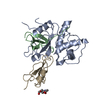
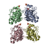
| Deposited unit | 
| ||||||||
|---|---|---|---|---|---|---|---|---|---|
| 1 | 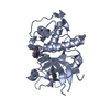
| ||||||||
| 2 | 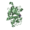
| ||||||||
| 3 | 
| ||||||||
| 4 | 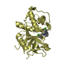
| ||||||||
| Unit cell |
|
-Components
| #1: Protein | / MAJOR EXCRETED PROTEIN / MEP Mass: 24161.676 Da / Num. of mol.: 4 / Fragment: CATALYTIC DOMAIN, RESIDUES 114-333 / Mutation: YES Source method: isolated from a genetically manipulated source Source: (gene. exp.) HOMO SAPIENS (human) / Cell line (production host): SF9 / Production host:   SPODOPTERA FRUGIPERDA (fall armyworm) / References: UniProt: P07711, cathepsin L SPODOPTERA FRUGIPERDA (fall armyworm) / References: UniProt: P07711, cathepsin L#2: Chemical | ChemComp-424 / (   Mass: 534.455 Da / Num. of mol.: 4 / Source method: obtained synthetically / Formula: C25H25Cl2N3O4S Mass: 534.455 Da / Num. of mol.: 4 / Source method: obtained synthetically / Formula: C25H25Cl2N3O4S#3: Water | ChemComp-HOH / | Water Mass: 18.015 Da / Num. of mol.: 720 / Source method: isolated from a natural source / Formula: H2O Mass: 18.015 Da / Num. of mol.: 720 / Source method: isolated from a natural source / Formula: H2ONonpolymer details | PROTEIN WAS CRYSTALLIZED AFTER TREATMENT WITH (2S, 4R)-4-(2-CHLORO-BENZENESULFONYL)-1-[1-(4-CHLORO- ...PROTEIN WAS CRYSTALLIZ | Sequence details | THR110 TO ALA MUTANT (T110A) CORRESPOND | |
|---|
-Experimental details
-Experiment
| Experiment | Method: X-RAY DIFFRACTION / Number of used crystals: 1 |
|---|
- Sample preparation
| Crystal | Density Matthews: 2.14 Å3/Da / Density % sol: 42.5 % / Description: ISOMORPHOUS TO PDB ENTRY 3HWN |
|---|---|
| Crystal grow | pH: 4 / Details: 20% PEG 2000, PH 4 |
-Data collection
| Diffraction | Mean temperature: 100 K |
|---|---|
| Diffraction source | Source: SYNCHROTRON / Site: SLS  / Beamline: X06DA / Wavelength: 1.008 / Beamline: X06DA / Wavelength: 1.008 |
| Detector | Type: MARMOSAIC 225 mm CCD / Detector: CCD / Date: Oct 7, 2009 |
| Radiation | Protocol: SINGLE WAVELENGTH / Monochromatic (M) / Laue (L): M / Scattering type: x-ray |
| Radiation wavelength | Wavelength: 1.008 Å / Relative weight: 1 |
| Reflection | Resolution: 1.45→50 Å / Num. obs: 137183 / % possible obs: 95.4 % / Observed criterion σ(I): -3 / Redundancy: 2.28 % / Biso Wilson estimate: 15.6 Å2 / Rmerge(I) obs: 0.12 / Net I/σ(I): 5.79 |
| Reflection shell | Resolution: 1.45→1.55 Å / Redundancy: 2.29 % / Rmerge(I) obs: 0.52 / Mean I/σ(I) obs: 1.46 / % possible all: 93.7 |
- Processing
| Software |
| ||||||||||||||||||||||||||||||||||||||||||||||||||||||||||||||||||||||||||||||||||||||||||||||||||||||||||||||||||||||||||||||||||||||||||||||||||||||||||||||||||||||||||||||||||||||
|---|---|---|---|---|---|---|---|---|---|---|---|---|---|---|---|---|---|---|---|---|---|---|---|---|---|---|---|---|---|---|---|---|---|---|---|---|---|---|---|---|---|---|---|---|---|---|---|---|---|---|---|---|---|---|---|---|---|---|---|---|---|---|---|---|---|---|---|---|---|---|---|---|---|---|---|---|---|---|---|---|---|---|---|---|---|---|---|---|---|---|---|---|---|---|---|---|---|---|---|---|---|---|---|---|---|---|---|---|---|---|---|---|---|---|---|---|---|---|---|---|---|---|---|---|---|---|---|---|---|---|---|---|---|---|---|---|---|---|---|---|---|---|---|---|---|---|---|---|---|---|---|---|---|---|---|---|---|---|---|---|---|---|---|---|---|---|---|---|---|---|---|---|---|---|---|---|---|---|---|---|---|---|---|
| Refinement | Method to determine structure: OTHER Starting model: NONE Resolution: 1.45→36.52 Å / Cor.coef. Fo:Fc: 0.916 / Cor.coef. Fo:Fc free: 0.891 / SU B: 2.292 / SU ML: 0.086 / Cross valid method: THROUGHOUT / ESU R: 0.105 / ESU R Free: 0.106 / Stereochemistry target values: MAXIMUM LIKELIHOOD / Details: HYDROGENS HAVE BEEN ADDED IN THE RIDING POSITIONS.
| ||||||||||||||||||||||||||||||||||||||||||||||||||||||||||||||||||||||||||||||||||||||||||||||||||||||||||||||||||||||||||||||||||||||||||||||||||||||||||||||||||||||||||||||||||||||
| Solvent computation | Ion probe radii: 0.8 Å / Shrinkage radii: 0.8 Å / VDW probe radii: 1.2 Å / Solvent model: BABINET MODEL WITH MASK | ||||||||||||||||||||||||||||||||||||||||||||||||||||||||||||||||||||||||||||||||||||||||||||||||||||||||||||||||||||||||||||||||||||||||||||||||||||||||||||||||||||||||||||||||||||||
| Displacement parameters | Biso mean: 11.311 Å2
| ||||||||||||||||||||||||||||||||||||||||||||||||||||||||||||||||||||||||||||||||||||||||||||||||||||||||||||||||||||||||||||||||||||||||||||||||||||||||||||||||||||||||||||||||||||||
| Refinement step | Cycle: LAST / Resolution: 1.45→36.52 Å
| ||||||||||||||||||||||||||||||||||||||||||||||||||||||||||||||||||||||||||||||||||||||||||||||||||||||||||||||||||||||||||||||||||||||||||||||||||||||||||||||||||||||||||||||||||||||
| Refine LS restraints |
|